Wang H et al. (JAN 2012)
Journal of translational medicine 10 1 167
Oncolytic vaccinia virus GLV-1h68 strain shows enhanced replication in human breast cancer stem-like cells in comparison to breast cancer cells.
BACKGROUND: Recent data suggest that cancer stem cells (CSCs) play an important role in cancer,as these cells possess enhanced tumor-forming capabilities and are responsible for relapses after apparently curative therapies have been undertaken. Hence,novel cancer therapies will be needed to test for both tumor regression and CSC targeting. The use of oncolytic vaccinia virus (VACV) represents an attractive anti-tumor approach and is currently under evaluation in clinical trials. The purpose of this study was to demonstrate whether VACV does kill CSCs that are resistant to irradiation and chemotherapy. METHODS: Cancer stem-like cells were identified and separated from the human breast cancer cell line GI-101A by virtue of increased aldehyde dehydrogenase 1 (ALDH1) activity as assessed by the ALDEFLUOR assay and cancer stem cell-like features such as chemo-resistance,irradiation-resistance and tumor-initiating were confirmed in cell culture and in animal models. VACV treatments were applied to both ALDEFLUOR-positive cells in cell culture and in xenograft tumors derived from these cells. Moreover,we identified and isolated CD44(+)CD24(+)ESA(+) cells from GI-101A upon an epithelial-mesenchymal transition (EMT). These cells were similarly characterized both in cell culture and in animal models. RESULTS: We demonstrated for the first time that the oncolytic VACV GLV-1h68 strain replicated more efficiently in cells with higher ALDH1 activity that possessed stem cell-like features than in cells with lower ALDH1 activity. GLV-1h68 selectively colonized and eventually eradicated xenograft tumors originating from cells with higher ALDH1 activity. Furthermore,GLV-1h68 also showed preferential replication in CD44(+)CD24(+)ESA(+) cells derived from GI-101A upon an EMT induction as well as in xenograft tumors originating from these cells that were more tumorigenic than CD44(+)CD24(-)ESA(+) cells. CONCLUSIONS: Taken together,our findings indicate that GLV-1h68 efficiently replicates and kills cancer stem-like cells. Thus,GLV-1h68 may become a promising agent for eradicating both primary and metastatic tumors,especially tumors harboring cancer stem-like cells that are resistant to chemo and/or radiotherapy and may be responsible for recurrence of tumors.
View Publication
产品号#:
01700
01705
05620
01702
产品名:
ALDEFLUOR™ 试剂盒
ALDEFLUOR™ DEAB试剂, 1.5 mM, 1 mL
MammoCult™ 人源培养基套装
ALDEFLUOR™检测缓冲液
Kaur R et al. (OCT 2015)
Disease models & mechanisms 8 10 1295--1309
OTX2 exhibits cell-context-dependent effects on cellular and molecular properties of human embryonic neural precursors and medulloblastoma cells.
Medulloblastoma (MB) is the most common malignant primary pediatric brain tumor and is currently divided into four subtypes based on different genomic alterations,gene expression profiles and response to treatment: WNT,Sonic Hedgehog (SHH),Group 3 and Group 4. This extensive heterogeneity has made it difficult to assess the functional relevance of genes to malignant progression. For example,expression of the transcription factor Orthodenticle homeobox2 (OTX2) is frequently dysregulated in multiple MB variants; however,its role may be subtype specific. We recently demonstrated that neural precursors derived from transformed human embryonic stem cells (trans-hENs),but not their normal counterparts (hENs),resemble Groups 3 and 4 MB in vitro and in vivo. Here,we tested the utility of this model system as a means of dissecting the role of OTX2 in MB using gain- and loss-of-function studies in hENs and trans-hENs,respectively. Parallel experiments with MB cells revealed that OTX2 exerts inhibitory effects on hEN and SHH MB cells by regulating growth,self-renewal and migration in vitro and tumor growth in vivo. This was accompanied by decreased expression of pluripotent genes,such as SOX2,and was supported by overexpression of SOX2 in OTX2+ SHH MB and hENs that resulted in significant rescue of self-renewal and cell migration. By contrast,OTX2 is oncogenic and promotes self-renewal of trans-hENs and Groups 3 and 4 MB independent of pluripotent gene expression. Our results demonstrate a novel role for OTX2 in self-renewal and migration of hENs and MB cells and reveal a cell-context-dependent link between OTX2 and pluripotent genes. Our study underscores the value of human embryonic stem cell derivatives as alternatives to cell lines and heterogeneous patient samples for investigating the contribution of key developmental regulators to MB progression.
View Publication
产品号#:
05850
05857
05870
05875
85850
85857
85870
85875
产品名:
mTeSR™1
mTeSR™1
Sancho-Martinez I et al. (FEB 2016)
Nature communications 7 10743
Establishment of human iPSC-based models for the study and targeting of glioma initiating cells.
Glioma tumour-initiating cells (GTICs) can originate upon the transformation of neural progenitor cells (NPCs). Studies on GTICs have focused on primary tumours from which GTICs could be isolated and the use of human embryonic material. Recently,the somatic genomic landscape of human gliomas has been reported. RTK (receptor tyrosine kinase) and p53 signalling were found dysregulated in ∼90% and 86% of all primary tumours analysed,respectively. Here we report on the use of human-induced pluripotent stem cells (hiPSCs) for modelling gliomagenesis. Dysregulation of RTK and p53 signalling in hiPSC-derived NPCs (iNPCs) recapitulates GTIC properties in vitro. In vivo transplantation of transformed iNPCs leads to highly aggressive tumours containing undifferentiated stem cells and their differentiated derivatives. Metabolic modulation compromises GTIC viability. Last,screening of 101 anti-cancer compounds identifies three molecules specifically targeting transformed iNPCs and primary GTICs. Together,our results highlight the potential of hiPSCs for studying human tumourigenesis.
View Publication
产品号#:
05850
05857
05870
05875
85850
85857
85870
85875
产品名:
mTeSR™1
mTeSR™1
Vanden Bempt M et al. (MAR 2016)
Leukemia March 8 Epub ahead of print
Generation of the Fip1l1–Pdgfra fusion gene using CRISPR/Cas genome editing
Krishnamurthy S et al. (DEC 2010)
Cancer research 70 23 9969--78
Endothelial cell-initiated signaling promotes the survival and self-renewal of cancer stem cells.
Recent studies have demonstrated that cancer stem cells play an important role in the pathobiology of head and neck squamous cell carcinomas (HNSCC). However,little is known about functional interactions between head and neck cancer stem-like cells (CSC) and surrounding stromal cells. Here,we used aldehyde dehydrogenase activity and CD44 expression to sort putative stem cells from primary human HNSCC. Implantation of 1,000 CSC (ALDH+CD44+Lin-) led to tumors in 13 (out of 15) mice,whereas 10,000 noncancer stem cells (ALDH-CD44-Lin-) resulted in 2 tumors in 15 mice. These data demonstrated that ALDH and CD44 select a subpopulation of cells that are highly tumorigenic. The ability to self-renew was confirmed by the observation that ALDH+CD44+Lin- cells sorted from human HNSCC formed more spheroids (orospheres) in 3-D agarose matrices or ultra-low attachment plates than controls and were serially passaged in vivo. We observed that approximately 80% of the CSC were located in close proximity (within 100-μm radius) of blood vessels in human tumors,suggesting the existence of perivascular niches in HNSCC. In vitro studies demonstrated that endothelial cell-secreted factors promoted self-renewal of CSC,as demonstrated by the upregulation of Bmi-1 expression and the increase in the number of orospheres as compared with controls. Notably,selective ablation of tumor-associated endothelial cells stably transduced with a caspase-based artificial death switch (iCaspase-9) caused a marked reduction in the fraction of CSC in xenograft tumors. Collectively,these findings indicate that endothelial cell-initiated signaling can enhance the survival and self-renewal of head and neck CSC.
View Publication
产品号#:
01700
01705
01702
产品名:
ALDEFLUOR™ 试剂盒
ALDEFLUOR™ DEAB试剂, 1.5 mM, 1 mL
ALDEFLUOR™检测缓冲液
Casazza A et al. (APR 2011)
Arteriosclerosis,thrombosis,and vascular biology 31 4 741--9
Systemic and targeted delivery of semaphorin 3A inhibits tumor angiogenesis and progression in mouse tumor models.
OBJECTIVE: The role of semaphorins in tumor progression is still poorly understood. In this study,we aimed at elucidating the regulatory role of semaphorin 3A (SEMA3A) in primary tumor growth and metastatic dissemination. METHODS AND RESULTS: We used 3 different experimental approaches in mouse tumor models: (1) overexpression of SEMA3A in tumor cells,(2) systemic expression of SEMA3A following liver gene transfer in mice,and (3) tumor-targeted release of SEMA3A using gene modified Tie2-expressing monocytes as delivery vehicles. In each of these experimental settings,SEMA3A efficiently inhibited tumor growth by inhibiting vessel function and increasing tumor hypoxia and necrosis,without promoting metastasis. We further show that the expression of the receptor neuropilin-1 in tumor cells is required for SEMA3A-dependent inhibition of tumor cell migration in vitro and metastatic spreading in vivo. CONCLUSIONS: In sum,both systemic and tumor-targeted delivery of SEMA3A inhibits tumor angiogenesis and tumor growth in multiple mouse models; moreover,SEMA3A inhibits the metastatic spreading from primary tumors. These data support the rationale for further investigation of SEMA3A as an anticancer molecule.
View Publication
产品号#:
09600
09650
19756
19756RF
产品名:
StemSpan™ SFEM
StemSpan™ SFEM
Kryczek I et al. (JAN 2012)
International journal of cancer. Journal international du cancer 130 1 29--39
Expression of aldehyde dehydrogenase and CD133 defines ovarian cancer stem cells.
Identification of cancer stem cells is crucial for advancing cancer biology and therapy. Several markers including CD24,CD44,CD117,CD133,the G subfamily of ATP-binding cassette transporters (ABCG),epithelial specific antigen (ESA) and aldehyde dehydrogenase (ALDH) are used to identify and investigate human epithelial cancer stem cells in the literature. We have now systemically analyzed and compared the expression of these markers in fresh ovarian epithelial carcinomas. Although the expression levels of these markers were unexpectedly variable and partially overlapping in fresh ovarian cancer cells from different donors,we reliably detected important levels of CD133 and ALDH in the majority of fresh ovarian cancer. Furthermore,most of these stem cell markers including CD133 and ALDH were gradually lost following in vitro passage of primary tumor cells. However,the expression of ALDH and CD133,but not CD24,CD44 and CD117,could be partially rescued by the in vitro serum-free and sphere cultures and by the in vivo passage in the immune-deficient xenografts. ALDH+ and CD133+ cells formed three-dimensional spheres more efficiently than their negative counterparts. These sphere-forming cells expressed high levels of stem cell core gene transcripts and could be expanded and form additional spheres in long-term culture. ALDH+,CD133+ and ALDH+ CD133+ cells from fresh tumors developed larger tumors more rapidly than their negative counterparts. This property was preserved in the xenografted tumors. Altogether,the data suggest that ALDH+ and CD133+ cells are enriched with ovarian cancer-initiating (stem) cells and that ALDH and CD133 may be widely used as reliable markers to investigate ovarian cancer stem cell biology.
View Publication
产品号#:
01700
01705
01701
01702
18555
18555RF
18551
18551RF
18561
产品名:
ALDEFLUOR™ 试剂盒
ALDEFLUOR™ DEAB试剂, 1.5 mM, 1 mL
ALDEFLUOR™检测缓冲液
Silva IA et al. (JUN 2011)
Cancer research 71 11 3991--4001
Aldehyde dehydrogenase in combination with CD133 defines angiogenic ovarian cancer stem cells that portend poor patient survival.
Markers that reliably identify cancer stem cells (CSC) in ovarian cancer could assist prognosis and improve strategies for therapy. CD133 is a reported marker of ovarian CSC. Aldehyde dehydrogenase (ALDH) activity is a reported CSC marker in several solid tumors,but it has not been studied in ovarian CSC. Here we report that dual positivity of CD133 and ALDH defines a compelling marker set in ovarian CSC. All human ovarian tumors and cell lines displayed ALDH activity. ALDH(+) cells isolated from ovarian cancer cell lines were chemoresistant and preferentially grew tumors,compared with ALDH(-) cells,validating ALDH as a marker of ovarian CSC in cell lines. Notably,as few as 1,000 ALDH(+) cells isolated directly from CD133(-) human ovarian tumors were sufficient to generate tumors in immunocompromised mice,whereas 50,000 ALDH(-) cells were unable to initiate tumors. Using ALDH in combination with CD133 to analyze ovarian cancer cell lines,we observed even greater growth in the ALDH(+)CD133(+) cells compared with ALDH(+)CD133(-) cells,suggesting a further enrichment of ovarian CSC in ALDH(+)CD133(+) cells. Strikingly,as few as 11 ALDH(+)CD133(+) cells isolated directly from human tumors were sufficient to initiate tumors in mice. Like other CSC,ovarian CSC exhibited increased angiogenic capacity compared with bulk tumor cells. Finally,the presence of ALDH(+)CD133(+) cells in debulked primary tumor specimens correlated with reduced disease-free and overall survival in ovarian cancer patients. Taken together,our findings define ALDH and CD133 as a functionally significant set of markers to identify ovarian CSCs.
View Publication
Noninvasive MR imaging of magnetically labeled stem cells to directly identify neovasculature in a glioma model.
Bone marrow-derived endothelial precursor cells incorporate into neovasculature and have been successfully used as vehicles for gene delivery to brain tumors. To determine whether systemically administered Sca1+ bone marrow cells labeled with superparamagnetic iron oxide nanoparticles can be detected by in vivo magnetic resonance imaging in a mouse brain tumor model,mouse Sca1+ cells were labeled in vitro with ferumoxides-poly-L-lysine complexes. Labeled or control cells were administered intravenously to glioma-bearing severe combined immunodeficient (SCID) mice. Magnetic resonance imaging (MRI) was performed during tumor growth. Mice that received labeled cells demonstrated hypointense regions within the tumor that evolved over time and developed a continuous dark hypointense ring at a consistent time point. This effect was not cleared by administration of a gadolinium contrast agent. Histology showed iron-labeled cells around the tumor rim in labeled mice,which expressed CD31 and von Willebrand factor,indicating the transplanted cells detected in the tumor have differentiated into endothelial-like cells. These results demonstrate that MRI can detect the incorporation of magnetically labeled bone marrow-derived precursor cells into tumor vasculature as part of ongoing angiogenesis and neovascularization. This technique can be used to directly identify neovasculature in vivo and to facilitate gene therapy by noninvasively monitoring these cells as gene delivery vectors.
View Publication
产品号#:
09600
09650
09850
产品名:
StemSpan™ SFEM
StemSpan™ SFEM
Kuroki MM et al. ( 2005)
Anticancer Research 25 6A 3733--9
Preparation of human IgG and IgM monoclonal antibodies for MK-1/Ep-CAM by using human immunoglobulin gene-transferred mouse and gene cloning of their variable regions.
For antibody-based therapy of cancer,monoclonal antibodies (mAbs) of human origin are superior to mouse,mouse/human chimeric or humanized mAbs,because of their minimum immunogenicity to humans and their efficient collaboration with human effector cells. In the present study,human mAbs were prepared against a pancarcinoma antigen,MK-1 (Ep-CAM),using a genetically-engineered mouse (KM mouse) that contains the human immunoglobulin genes. Spleen cells from KM mice,immunized with recombinant MK-1,were fused with P3-U1 mouse myeloma cells. Of 44 anti-MK-1 clones analyzed,two were of IgG4 and the others of IgM clones. Although the two IgG4 clones were suggested to recognize the same antigenic determinant or two closely located determinants,their VK regions were encoded by different light-chain genes while their VH sequences were identical. The two IgG4 and one of the IgM clones tested revealed antibody-dependent cell-mediated cytotoxicity and complement-dependent cytotoxicity,respectively,against MK-1-expressing cells in vitro,suggesting that these fully human mAbs produced against MK-1 and their V-region genes,which are applicable for the preparation of engineered antibody fragments that may be useful for antibody-based therapy of cancer.
View Publication

 EasySep™小鼠TIL(CD45)正选试剂盒


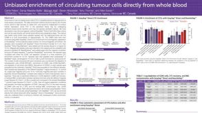 科学海报Unbiased Enrichment of Circulating Tumour Cells Directly from Whole Blood

 沪公网安备31010102008431号