Loss of p19Arf in a Rag1(-/-) B-cell precursor population initiates acute B-lymphoblastic leukemia.
In human B-acute lymphoblastic leukemia (B-ALL),RAG1-induced genomic alterations are important for disease progression. However,given that biallelic loss of the RAG1 locus is observed in a subset of cases,RAG1's role in the development of B-ALL remains unclear. We chose a p19Arf(-/-)Rag1(-/-) mouse model to confirm the previously published results concerning the contribution of CDKN2A (p19ARF /INK4a) and RAG1 copy number alterations in precursor B cells to the initiation and/or progression to B-acute lymphoblastic leukemia (B-ALL). In this murine model,we identified a new,Rag1-independent leukemia-initiating mechanism originating from a Sca1(+)CD19(+) precursor cell population and showed that Notch1 expression accelerates the cells' self-renewal capacity in vitro. In human RAG1-deficient BM,a similar CD34(+)CD19(+) population expressed p19ARF. These findings suggest that combined loss of p19Arf and Rag1 results in B-cell precursor leukemia in mice and may contribute to the progression of precursor B-ALL in humans.
View Publication
产品号#:
产品名:
Zetterblad J et al. (JAN 2010)
BMC genomics 11 108
Genomics based analysis of interactions between developing B-lymphocytes and stromal cells reveal complex interactions and two-way communication.
BACKGROUND: The use of functional genomics has largely increased our understanding of cell biology and promises to help the development of systems biology needed to understand the complex order of events that regulates cellular differentiation in vivo. One model system clearly dependent on the integration of extra and intra cellular signals is the development of B-lymphocytes from hematopoietic stem cells in the bone marrow. This developmental pathway involves several defined differentiation stages associated with specific expression of genes including surface markers that can be used for the prospective isolation of the progenitor cells directly from the bone marrow to allow for ex vivo gene expression analysis. The developmental process can be simulated in vitro making it possible to dissect information about cell/cell communication as well as to address the relevance of communication pathways in a rather direct manner. Thus we believe that B-lymphocyte development represents a useful model system to take the first steps towards systems biology investigations in the bone marrow. RESULTS: In order to identify extra cellular signals that promote B lymphocyte development we created a database with approximately 400 receptor ligand pairs and software matching gene expression data from two cell populations to obtain information about possible communication pathways. Using this database and gene expression data from NIH3T3 cells (unable to support B cell development),OP-9 cells (strongly supportive of B cell development),pro-B and pre-B cells as well as mature peripheral B-lineage cells,we were able to identify a set of potential stage and stromal cell restricted communication pathways. Functional analysis of some of these potential ways of communication allowed us to identify BMP-4 as a potent stimulator of B-cell development in vitro. Further,the analysis suggested that there existed possibilities for progenitor B cells to send signals to the stroma. The functional consequences of this were investigated by co-culture experiments revealing that the co-incubation of stromal cells with B cell progenitors altered both the morphology and the gene expression pattern in the stromal cells. CONCLUSIONS: We believe that this gene expression data analysis method allows for the identification of functionally relevant interactions and therefore could be applied to other data sets to unravel novel communication pathways.
View Publication
产品号#:
产品名:
Staton PJ et al. (APR 2006)
Journal of immunology (Baltimore,Md. : 1950) 176 7 3978--86
IL-7 is a critical factor in modulating lesion development in Skn-directed autoimmunity.
In a murine model of autoimmunity targeted against the epidermal cell Ags,Skn,adoptive transfer of Skn-immune T cells to immunosuppressed recipients elicits skin lesions in areas of mild epidermal trauma. In this study,we examined peripheral regulation of Skn-induced autoreactivity disrupted by rendering the mice immunoincompetent. We found that regulation of Skn-directed autoimmunity was restored by cotransfer of normal syngeneic spleen cells at twice the concentration of Skn-immune cells and was evidenced by significantly reduced lesion severity by days 5-7 post-cotransfer compared with animals given injections of Skn-immune cells alone. Enrichment and depletion of normal CD4(+) or CD8(+) spleen cells and RT-PCR analysis of selected cytokines identified CD4(+) cells as the regulatory cells in the cotransfer inoculum; however,significant reduction in lesion severity was observed only when there was a concomitant increase in levels of IL-7. The role of IL-7 was further supported in that mice cotransferred with Skn-immune cells plus normal spleen cells,but also treated with anti-IL-7 Ab,no longer exhibited reduced lesion severity. To determine whether IL-7 expression without normal spleen cell cotransfer could modulate lesion development,an IL-7-encoding plasmid (pCMV-Tag1-IL-7) was topically delivered to sites flanking the stressed skin site in Skn-induced autoimmune mice. Daily application of 15 mug of pCMV-Tag1-IL-7 significantly suppressed lesion severity. Our results support a mechanism for CD4(+) T cells and IL-7 in contributing to the control of autoreactivity.
View Publication
产品号#:
产品名:
Le Y et al. (MAR 2005)
Journal of immunology (Baltimore,Md. : 1950) 174 5 2582--90
CXC chemokine ligand 12-induced focal adhesion kinase activation and segregation into membrane domains is modulated by regulator of G protein signaling 1 in pro-B cells.
CXCL12-induced chemotaxis and adhesion to VCAM-1 decrease as B cells differentiate in the bone marrow. However,the mechanisms that regulate CXCL12/CXCR4-mediated signaling are poorly understood. We report that after CXCL12 stimulation of progenitor B cells,focal adhesion kinase (FAK) and PI3K are inducibly recruited to raft-associated membrane domains. After CXCL12 stimulation,phosphorylated FAK is also localized in membrane domains. The CXCL12/CXCR4-FAK pathway is membrane cholesterol dependent and impaired by metabolic inhibitors of G(i),Src family,and the GTPase-activating protein,regulator of G protein signaling 1 (RGS1). In the bone marrow,RGS1 mRNA expression is low in progenitor B cells and high in mature B cells,implying developmental regulation of CXCL12/CXCR4 signaling by RGS1. CXCL12-induced chemotaxis and adhesion are impaired when FAK recruitment and phosphorylation are inhibited by either membrane cholesterol depletion or overexpression of RGS1 in progenitor B cells. We conclude that the recruitment of signaling molecules to specific membrane domains plays an important role in CXCL12/CXCR4-induced cellular responses.
View Publication
产品号#:
产品名:
Bruserud &O et al. (MAY 2003)
Leukemia research 27 5 455--64
In vitro culture of human acute lymphoblastic leukemia (ALL) cells in serum-free media; a comparison of native ALL blasts, ALL cell lines and virus-transformed B cell lines.
The aim of this study was to standardize in vitro culture conditions for human acute lymphoblastic leukemia (ALL) cells. The cells were cultured in medium containing 10% fetal calf serum (FCS) and in the four serum-free media X-vivo 10,X-vivo 15,X-vivo 20 and Stem Span. Native ALL blasts could proliferate in all four serum-free media,but the strongest responses were usually observed with Stem Span. Native leukemia blasts were also cultured in the presence of various single cytokines or cytokine combinations. The highest proliferation was usually observed in the presence of Flt3-Ligand (Flt3-L) when single cytokines were examined,and these responses could be further increased especially by combining Flt3-L with interleukin 3 (IL3),IL7 or stem cell factor (SCF). Proliferation could also be increased when ALL blasts were cultured in the presence of two commercially available fibroblast cell lines (Hs27 and HFL1). Based on these results we suggest that in vitro culture conditions for native human ALL blasts can be standardized by using serum-free culture media supplemented with exogenous Flt3-L+IL3+SCF,and the use of accessory cells can also be standardized by using well-characterized fibroblast cell lines. Detectable ALL blast proliferation can then be observed for most patients. Our experimental model can thereby be used for in vitro evaluation of possible antileukemic treatment strategies,and it will then allow comparison of experimental results between different studies.
View Publication

 EasySep™小鼠TIL(CD45)正选试剂盒


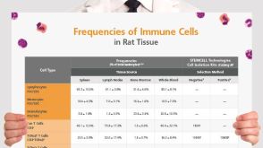 挂图Frequencies of Immune Cells in Rat Tissue Lists the estimated frequencies of more than 15 immune cell types in Sprague Dawley rats


 沪公网安备31010102008431号