
 EasySep™小鼠TIL(CD45)正选试剂盒


Scientific Resources
-
 技术公告Tissue Dissociation and Digestion Protocols
产品类型:
Tissue and Cell Culture Dissociation Reagents
-
-
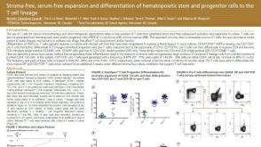 科学海报Stroma-Free, Serum-Free Expansion and Differentiation of Hematopoietic Stem and Progenitor Cells to the T Cell Lineage
产品类型:
Cell Culture Media and Supplements
Conference:
AAi 2017
-
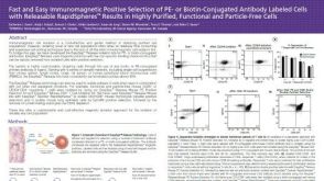 科学海报Positive Selection of PE- or Biotin-Conjugated Antibody Labeled Cells with Releasable Rapidspheres™
产品类型:
Cell Isolation Products
Conference:
AAI 2017


 沪公网安备31010102008431号