
 EasySep™小鼠TIL(CD45)正选试剂盒


产品号 #100-1365_C
无血清、无异种成分培养基,用于生成功能齐全、成熟的 hPSC 衍生视网膜色素上皮
若您需要咨询产品或有任何技术问题,请通过官方电话 400 885 9050 或邮箱 info.cn@stemcell.com 与我们联系。
无血清、无异种成分培养基,用于生成功能齐全、成熟的 hPSC 衍生视网膜色素上皮
无血清、无异种成分培养基,用于生成功能齐全、成熟的 hPSC 衍生视网膜色素上皮
无需手动筛选或细胞富集,即可快速成熟高纯度、功能性人多能干细胞 (hPSC) 衍生的视网膜色素上皮 (RPE) 细胞。STEMdiff™-XF RPE 成熟培养基是一种无异种成分、无血清培养基,支持在 35 天内将未成熟 RPE 分化为成熟且具有功能的 RPE。使用 STEMdiff™-XF RPE 成熟培养基生成的成熟 RPE 细胞表达成熟标志物(图 2),并展现出与原代 RPE 细胞相当的关键功能,包括色素沉着、多边形形态、极化和吞噬功能。本产品经过优化,可用于使用STEMdiff™-ACF RPE 分化试剂盒生成的 iPSC 衍生 RPE 细胞的成熟过程。STEMdiff™-XF RPE 成熟培养基 可与以下兼容的 STEMdiff™ RPE 产品组成一个完整且优化的工作流程:
STEMdiff™-ACF RPE 铺板补充剂
STEMdiff™-ACF RPE 分化试剂盒
如您计划将本产品用于商业或临床用途,请与我们联系。
细胞类型
多能干细胞
应用
细胞培养
品牌
STEMdiff
研究领域
疾病建模,药物发现和毒理检测,神经科学,细胞治疗开发
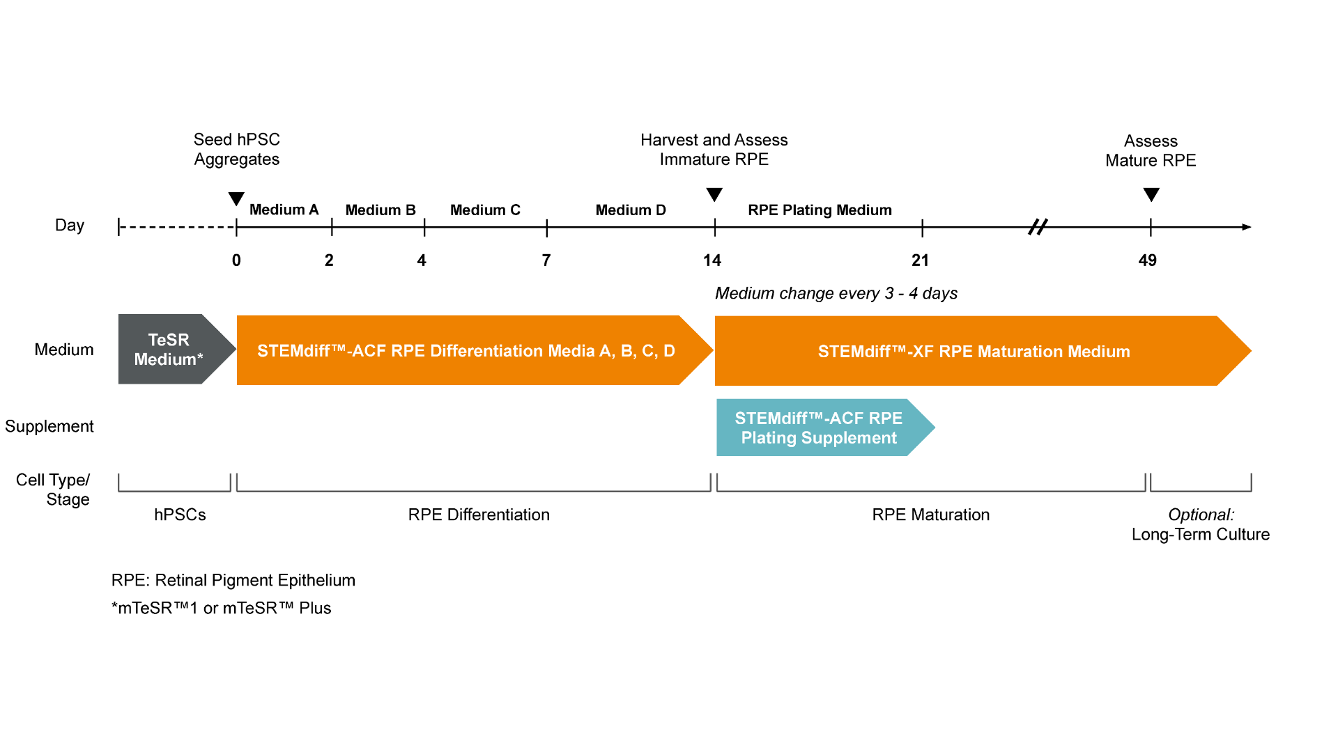
Figure 1. Workflow for the Differentiation of hPSCs into Retinal Pigment Epithelium (RPE) with STEMdiff™ RPE Culture System
hPSC colonies, previously harvested and seeded as clumps, are put directly into the medium provided in STEMdiff™-ACF RPE Differentiation Kit. Seeding into STEMdiff™ RPE Differentiation Medium A induces cells toward immature RPE. A full medium change is performed on Day 1 with fresh STEMdiff™ RPE Differentiation Medium A and then on Day 2 with STEMdiff™ RPE Differentiation Medium B. On Days 4 and 6, medium changes are performed with fresh STEMdiff™ RPE Differentiation Medium C. On Days 7 and every second day thereafter a medium change is performed with fresh STEMdiff™ RPE Differentiation Medium D. On Day 14, immature RPE cells are enzymatically harvested and subcultured in STEMdiff™-XF RPE Maturation Medium with the addition of STEMdiff™-ACF RPE Plating Supplement from Days 14 to 21 to improve survival after passaging. RPE cells begin to mature over the course of 5 weeks of culture in STEMdiff™-XF RPE Maturation Medium and become fully matured by Day 49, possessing key characteristics such as polygonal shape, polarization, pigmentation and phagocytosis.
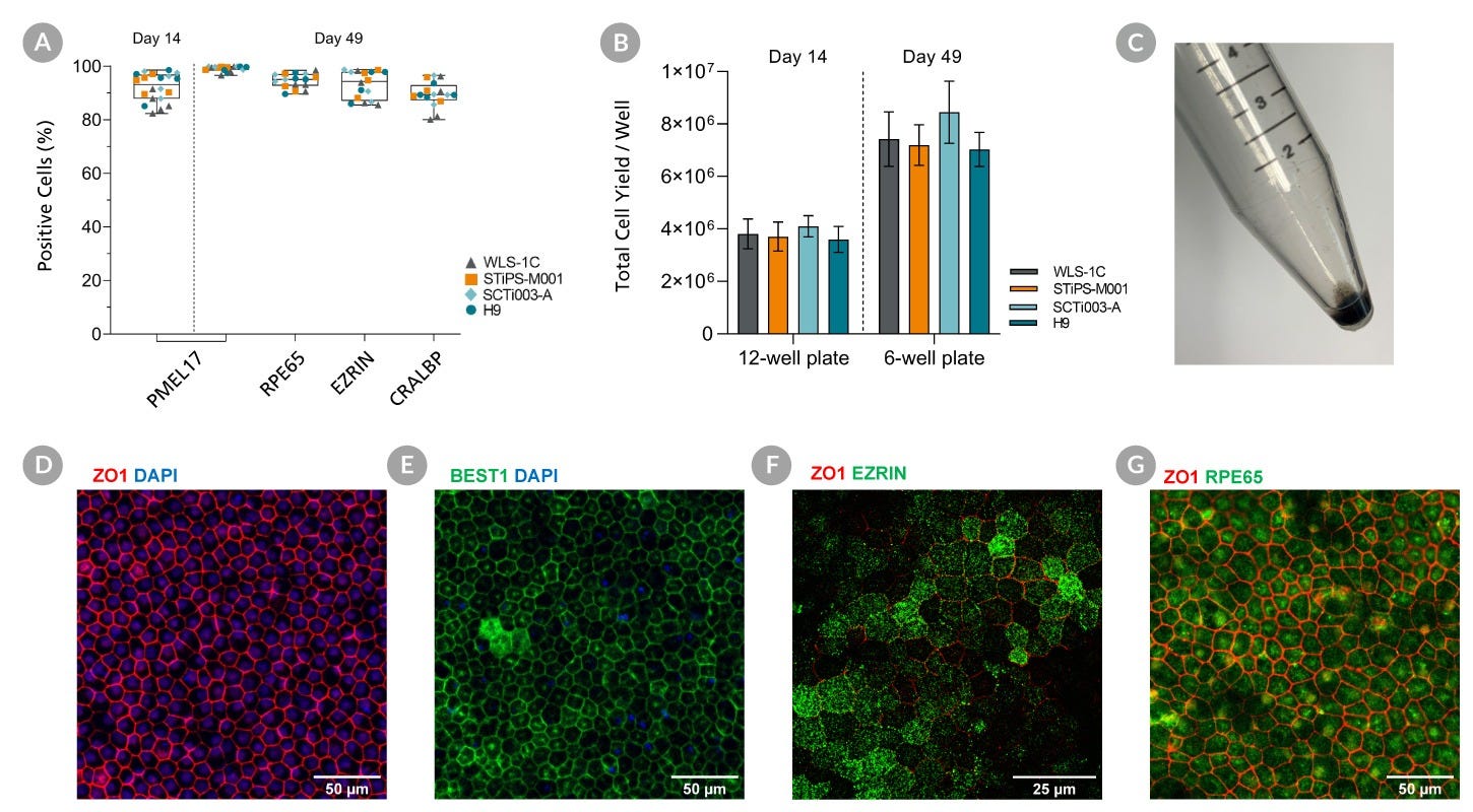
Figure 2. Robust and Rapid Generation of Mature Retinal Pigment Epithelial Cells (RPE) across multiple PSC Cell Lines with the STEMdiff™-ACF RPE Differentiation Kit
hPSCs were cultured for 14 Days using STEMdiff™-ACF RPE Differentiation Kit and subsequently subcultured in STEMdiff™-XF RPE Maturation Medium. Flow cytometry expression of RPE markers are shown at Day 14 and Day 49. (A) The percentage of cells expressing PMEL17, RPE65, EZRIN, and CRALBP and (B) Viable cell yields for 4 hPSC cell lines. PMEL17 is expressed at Day 14 and 49 while the other markers are only present at Day 49. Data are reported as mean + SEM; n = 16 -20. (C) A cell pellet of mature RPE cells demonstrates the pigmentation. Maturation is further demonstrated with immunohistochemistry for expression of RPE markers at Day 49. (D, E, F, G) Mature RPE display tight junctions marked by localization of ZO1 and BEST1 to cell junctions. Mature RPE are polarized, expressing EZRIN apically and ZO1 subapically and express proteins required for the visual cycle (RPE65).
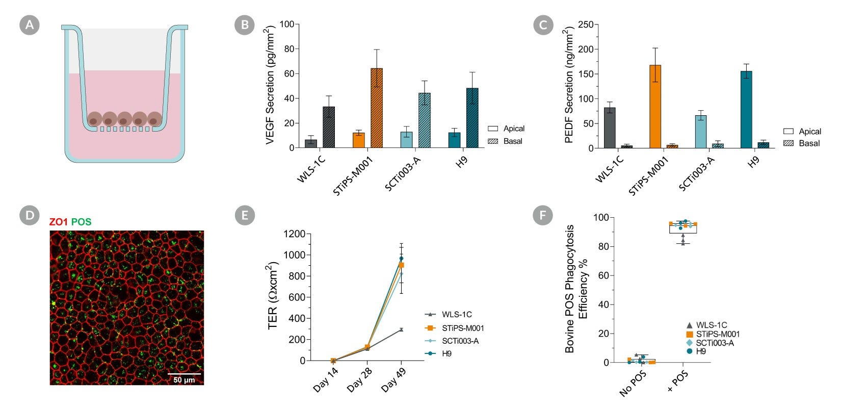
Figure 3. Mature Retinal Pigment Epithelial (RPE) Cells Display Key Functionalities Corresponding to RPE Behaviour
hPSC’s were cultured for 14 Days using STEMdiff™-ACF RPE Differentiation Kit and subsequently subcultured on cell culture inserts in STEMdiff™-XF RPE Maturation Medium for 5 weeks. Apical and basal conditioned medium were collected from Mature RPE, and a sandwich ELISA was performed to quantify Vascular Endothelial Growth Factor (VEGF) and Pigment Epithelial Derived Growth Factor (PEDF) secretion. (A, B) Mature RPE secreted more basal VEGF and apical PEDF demonstrating RPE display correct apicobasal polarity. Data shown as mean + SEM; n = 3. (C) Mature RPE were able to generate a strong barrier with high transepithelial resistance (TER). Data shown as mean + SEM; n = 3-6. (D) Mature RPE were fed FITC-labelled bovine photoreceptor outer segments (POS) for 4 to 5 hours prior to being enzymatically dissociated for flow cytometry analysis or fixed with paraformaldehyde for immunostaining. (E) Mature RPE efficiently internalize bovine POS. Data shown as mean + SEM; n = 3. (F) A cross-sectional schematic of the cell insert culture system.
请在《产品说明书》中查找相关支持信息和使用说明,或浏览下方更多实验方案。
本产品专为以下研究领域设计,适用于工作流程中的高亮阶段。探索这些工作流程,了解更多我们为各研究领域提供的其他配套产品。
Thank you for your interest in IntestiCult™ Organoid Growth Medium (Human). Please provide us with your contact information and your local representative will contact you with a customized quote. Where appropriate, they can also assist you with a(n):
Estimated delivery time for your area
Product sample or exclusive offer
In-lab demonstration
扫描二维码或搜索微信号STEMCELLTech,即可关注我们的微信平台,第一时间接收丰富的技术资源和最新的活动信息。
如您有任何问题,欢迎发消息给STEMCELLTech微信公众平台,或与我们通过电话/邮件联系:400 885 9050 INFO.CN@STEMCELL.COM。
在线联系

 沪公网安备31010102008431号