
 EasySep™小鼠TIL(CD45)正选试剂盒


产品号 #05795_C
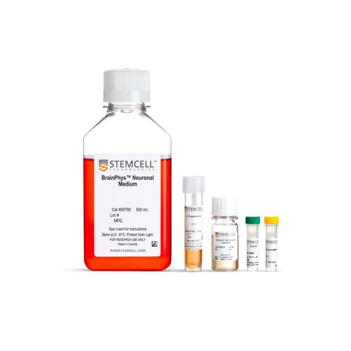
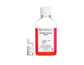
试剂盒包含 BrainPhys™ 神经元培养基、SM1 神经元补充剂、N2 补充剂-A、BDNF 和 GDNF,用于培养 ES/iPS 细胞衍生的神经元
若您需要咨询产品或有任何技术问题,请通过官方电话 400 885 9050 或邮箱 info.cn@stemcell.com 与我们联系。
试剂盒包含 BrainPhys™ 神经元培养基、SM1 神经元补充剂、N2 补充剂-A、BDNF 和 GDNF,用于培养 ES/iPS 细胞衍生的神经元
I want to help neuroscientists like you create more physiological culture conditions, for more active and healthy neuronal cultures.

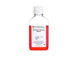
使用优化配方的完整培养基,可用于培养、分化并成熟由人胚胎干(ES)细胞或诱导多能干(iPS)细胞分化的神经元,其设计目的是促进而非抑制神经元活性。
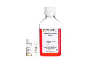
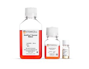
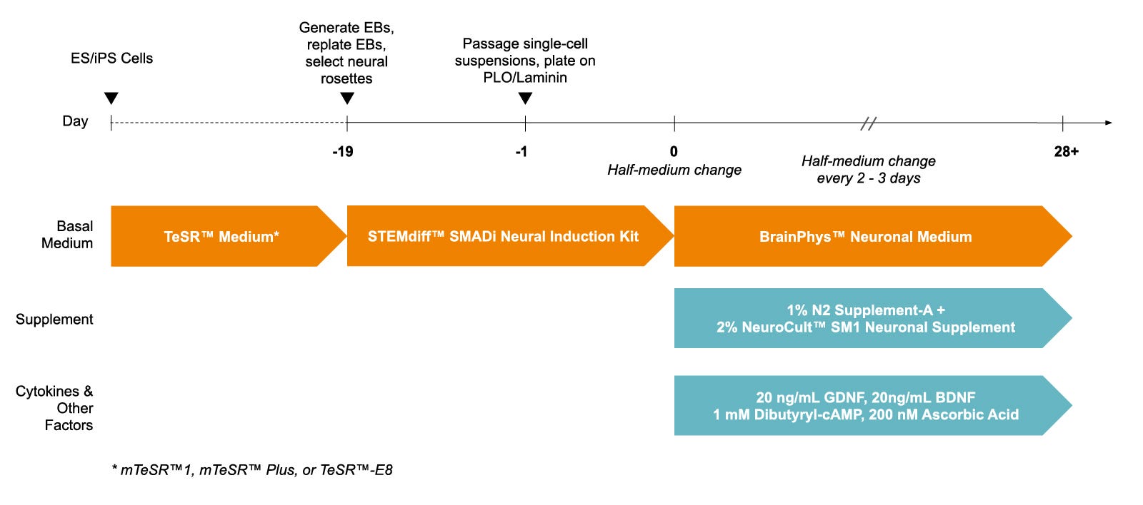
为提升使用便利性,BrainPhys™ hPSC神经元试剂盒提供了无血清BrainPhys™神经元培养基(基础培养基)、添加物和生长因子,帮助您从人ES/iPS细胞来源的神经祖细胞中生成并成熟不同类型的神经元。
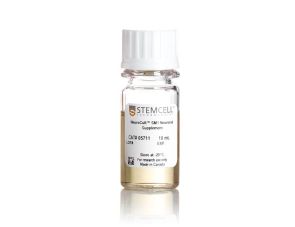
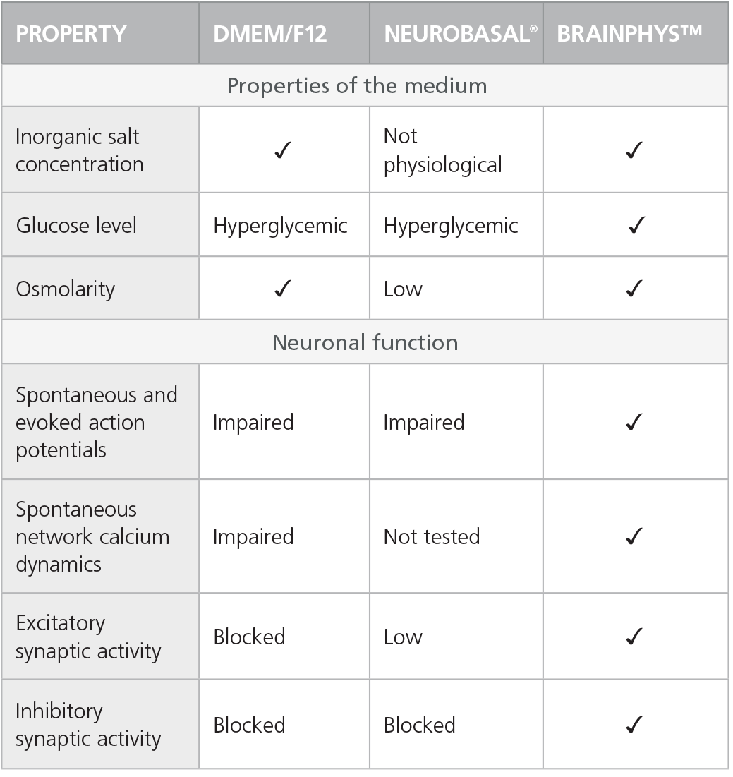
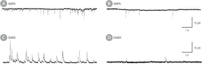
BrainPhys™ 神经元培养基基于 Bardy与Gage(Bardy et al. PNAS, 2015)的配方,模拟中枢神经系统(CNS)的细胞外环境,从而诱导更高比例的突触活性神经元。其中,基于Brewer B27配方(Brewer et al. J Neurosci Res., 1993)的NeuroCult™ SM1神经元添加物可维持细胞健康,并在短期与长期无血清培养条件下促进神经突起生长与分支;N2 Supplement-A则支持ES/iPS细胞向多种神经元亚型的分化。此外,试剂盒中还包含BDNF和GDNF生长因子,用于支持谱系特异性的分化。
为了避免因培养基更换而对细胞造成压力,您还可以在进行功能性分析(例如微电极阵列记录或实时荧光成像)时使用 BrainPhys™ 培养基。
查看我们的其他资源,了解更多关于BrainPhys™ 系统的信息。
分类
基础培养基,专用培养基
细胞类型
神经细胞,PSC衍生,神经元,多能干细胞
种属
人
应用
细胞培养,分化,培养
品牌
BrainPhys
研究领域
疾病建模,药物发现和毒理检测,神经科学,干细胞生物学
制剂类别
无血清
请在《产品说明书》中查找相关支持信息和使用说明,或浏览下方更多实验方案。
本产品专为以下研究领域设计,适用于工作流程中的高亮阶段。探索这些工作流程,了解更多我们为各研究领域提供的其他配套产品。
| 物种 | 人 |
|---|---|
| 配方 | 无血清 |
无血清神经添加物(50X)
提升神经元功能的无血清基础培养基
提升神经元功能的无血清基础培养基
试剂盒包括BrainPhys™神经元培养基和SM1神经元添加物,用于原代神经元以及胚胎干/诱导多能干细胞来源神经元的无血清培养
用于在BrainPhys™神经元培养基中无血清培养ES/iPS细胞来源的神经元
BrainPhys™ 原代神经元无血清培养试剂盒
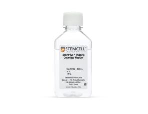
无血清神经生理基础培养基,改善神经元活细胞成像和功能

用于小鼠和人胚胎干细胞和iPS细胞的神经和胰腺分化
在线联系


 沪公网安备31010102008431号