
 EasySep™小鼠TIL(CD45)正选试剂盒


产品号 #05794_C
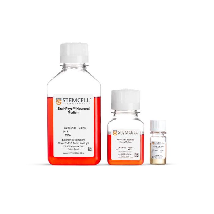
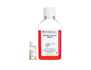
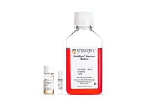
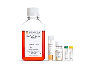
BrainPhys™ 原代神经元无血清培养试剂盒
若您需要咨询产品或有任何技术问题,请通过官方电话 400 885 9050 或邮箱 info.cn@stemcell.com 与我们联系。
BrainPhys™ 原代神经元无血清培养试剂盒
I want to help neuroscientists like you create more physiological culture conditions, for more active and healthy neuronal cultures.

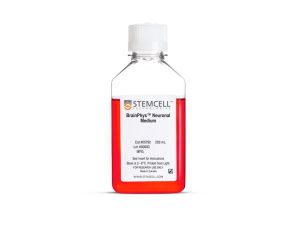
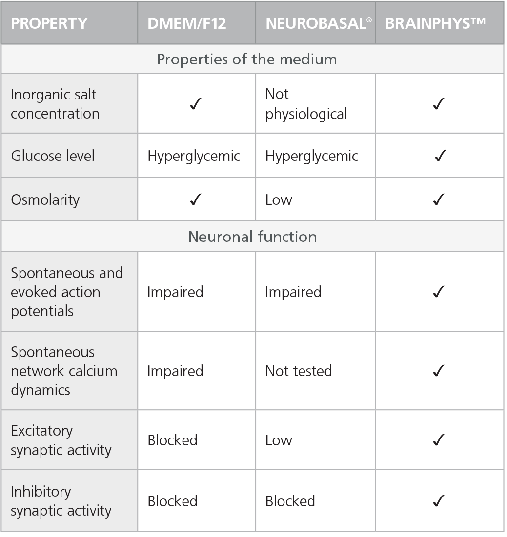
在完全无血清的原代神经元培养基中培养中枢神经系统 (CNS) 神经元,该培养基经过优化,可促进而非抑制神经元的活性和成熟。
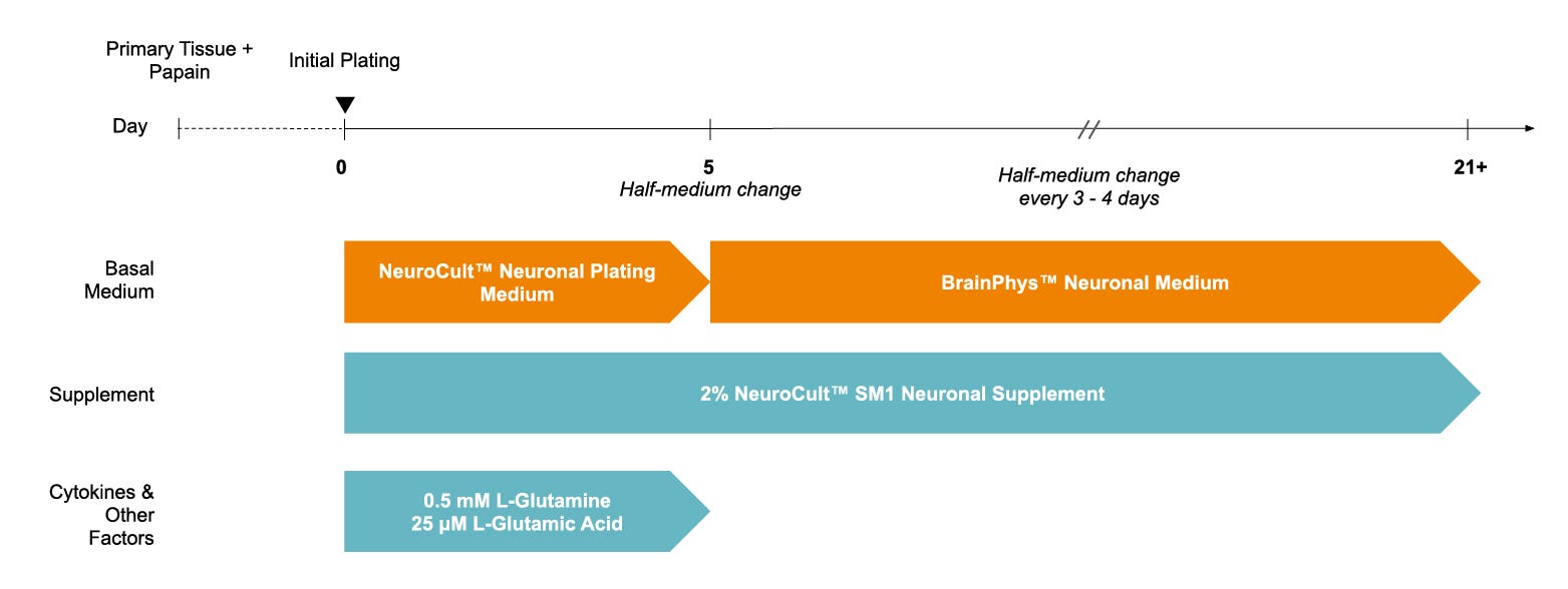
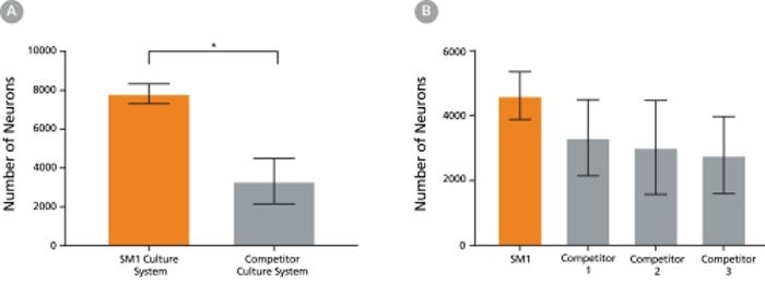
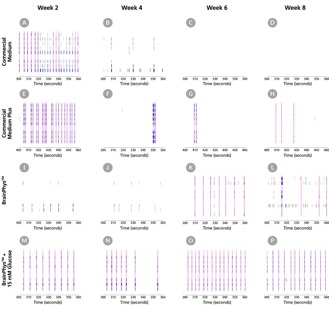
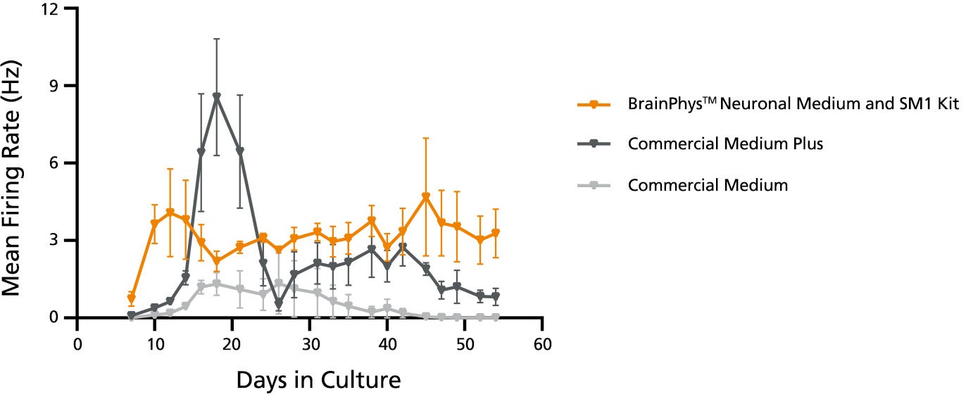
为了方便起见,BrainPhys™原代神经元培养试剂盒包含无血清的BrainPhys™神经元培养基(基础培养基)、NeuroCult™ SM1神经元添加物和NeuroCult™神经元铺板培养基,从而为您的原代组织来源神经元提供完整的培养体系。BrainPhys™神经元培养基基于Bardy和Gage的配方(Bardy et al. PNAS, 2015),可模拟中枢神经系统(CNS)的细胞外环境,以产生更高比例的突触活性神经元。NeuroCult™ SM1神经元添加物基于Brewer's B27配方(Brewer et al. J Neurosci Res., 1993),可在短期和长期无血清培养中维持细胞健康并促进神经突生长和分支。
为了避免因更换培养基而对细胞造成冲击,您也可以在进行功能性检测(例如基于微电极阵列的记录或活细胞荧光成像)时使用BrainPhys™培养基。
查看我们的其他资源,了解更多关于BrainPhys™产品线的信息。
分类
基础培养基,专用培养基
细胞类型
神经元
种属
人,小鼠,大鼠
应用
细胞培养,分化,培养
品牌
BrainPhys
研究领域
药物发现与毒性检测,神经科学,干细胞生物学
制剂类别
无血清
请在《产品说明书》中查找相关支持信息和使用说明,或浏览下方更多实验方案。
本产品专为以下研究领域设计,适用于工作流程中的高亮阶段。探索这些工作流程,了解更多我们为各研究领域提供的其他配套产品。
| 物种 | 人, 大鼠, 小鼠 |
|---|---|
| 配方 | 无血清 |
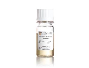
无血清神经添加物(50X)
提升神经元功能的无血清基础培养基
提升神经元功能的无血清基础培养基
试剂盒包括BrainPhys™神经元培养基和SM1神经元添加物,用于原代神经元以及胚胎干/诱导多能干细胞来源神经元的无血清培养
用于在BrainPhys™神经元培养基中无血清培养ES/iPS细胞来源的神经元
用于在BrainPhys™神经元培养基中无血清培养和分化ES/iPS细胞来源的神经元的试剂盒
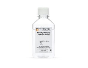
无血清神经生理基础培养基,改善神经元活细胞成像和功能

用于小鼠和人胚胎干细胞和iPS细胞的神经和胰腺分化
在线联系


 沪公网安备31010102008431号