
 EasySep™小鼠TIL(CD45)正选试剂盒


产品号 #200-0511_C
人多能干细胞系,冻存
若您需要咨询产品或有任何技术问题,请通过官方电话 400 885 9050 或邮箱 info.cn@stemcell.com 与我们联系。
人多能干细胞系,冻存
人多能干细胞系,冻存
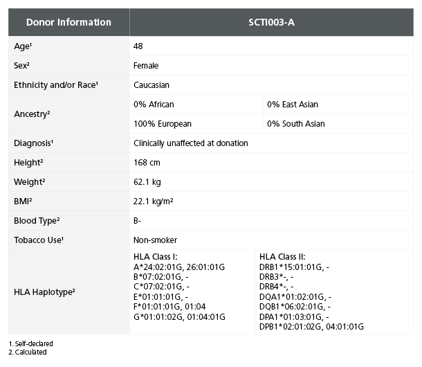
使用来自SCTi003-A细胞系的高质量诱导多能干细胞(iPSCs),自信地开始您的研究。SCTi003-A源自健康女性供体的外周血单个核细胞(PBMCs),适用于各种应用,例如用作未受影响的对照、基因编辑或向下游分化为谱系特异性细胞类型和类器官。
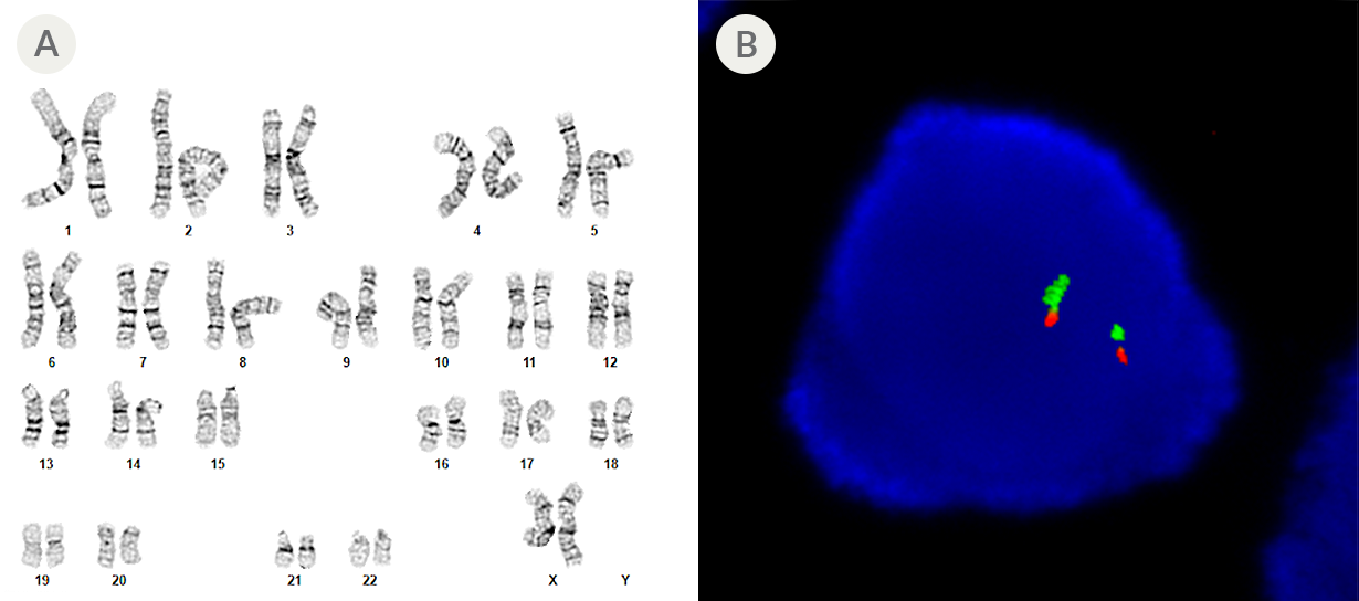
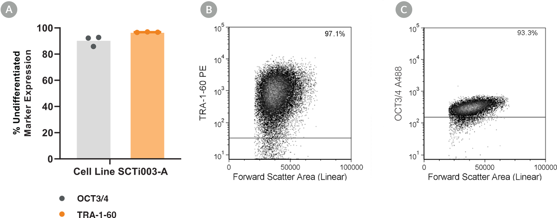
为确保最佳的产品性能和可重复性,SCTi003-A在包含mTeSR™ Plus、Corning® Matrigel® hESC-Qualified基质和ReLeSR™的培养系统中采用严格的质量控制程序生产。SCTi003-A具有稳定的核型,表现出三谱系分化潜能,表达未分化的细胞标志物,并使用非整合型重编程技术进行重编程。在hPSCreg®的注册确保了其基于社区标准的伦理和生物学一致性。
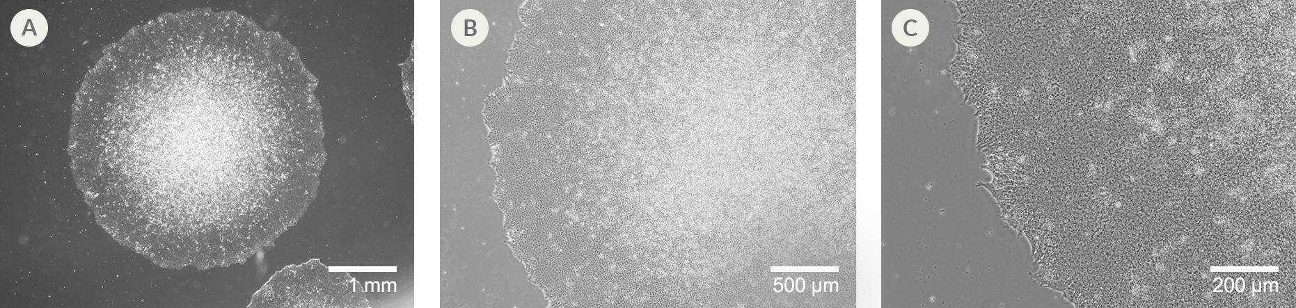
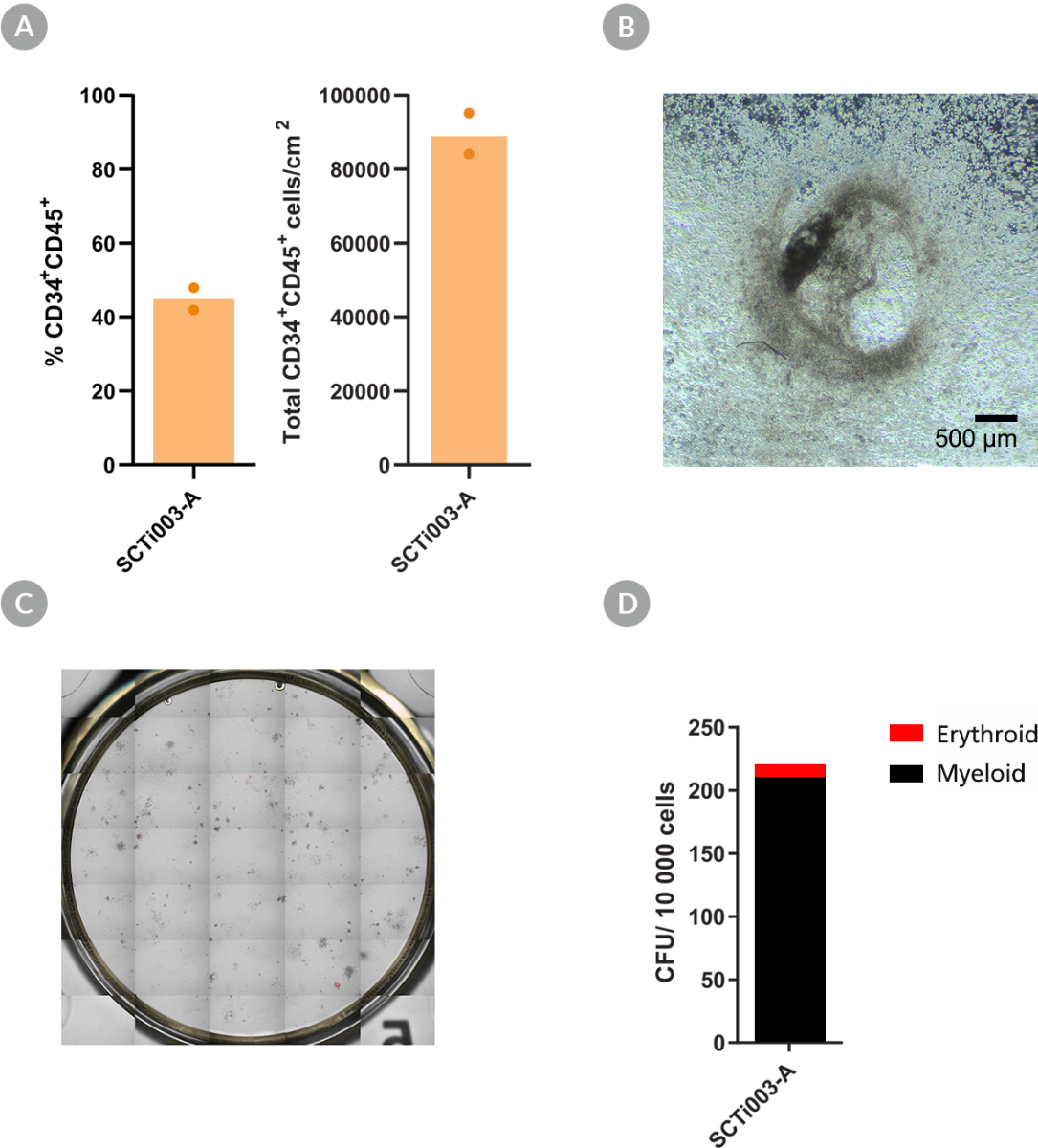
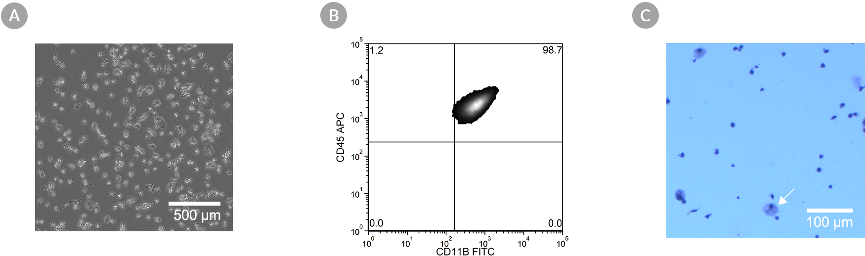
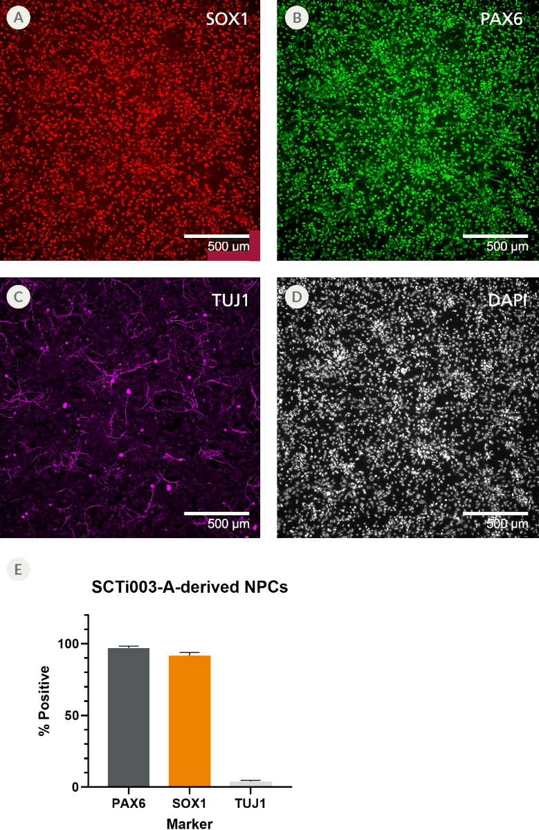
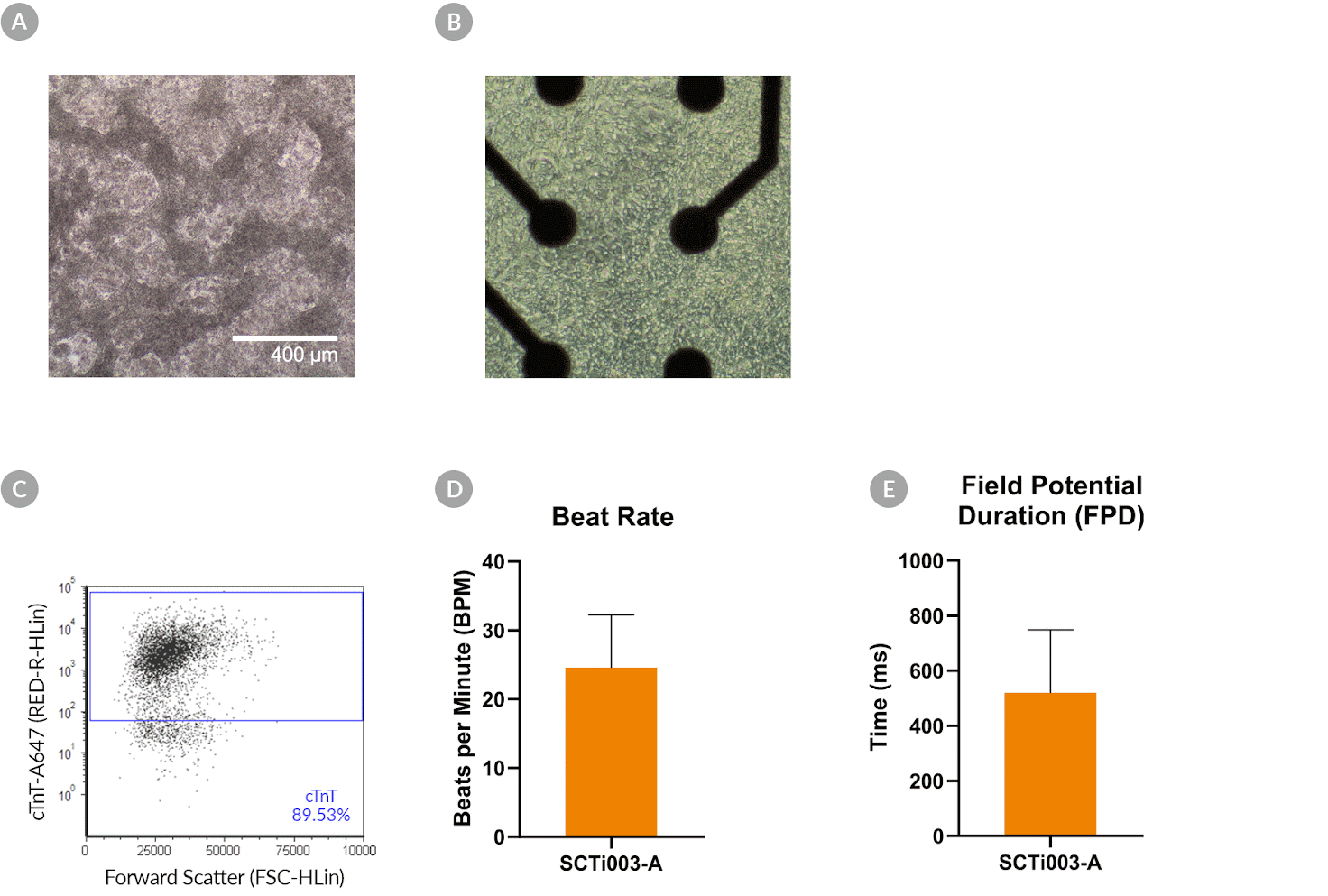
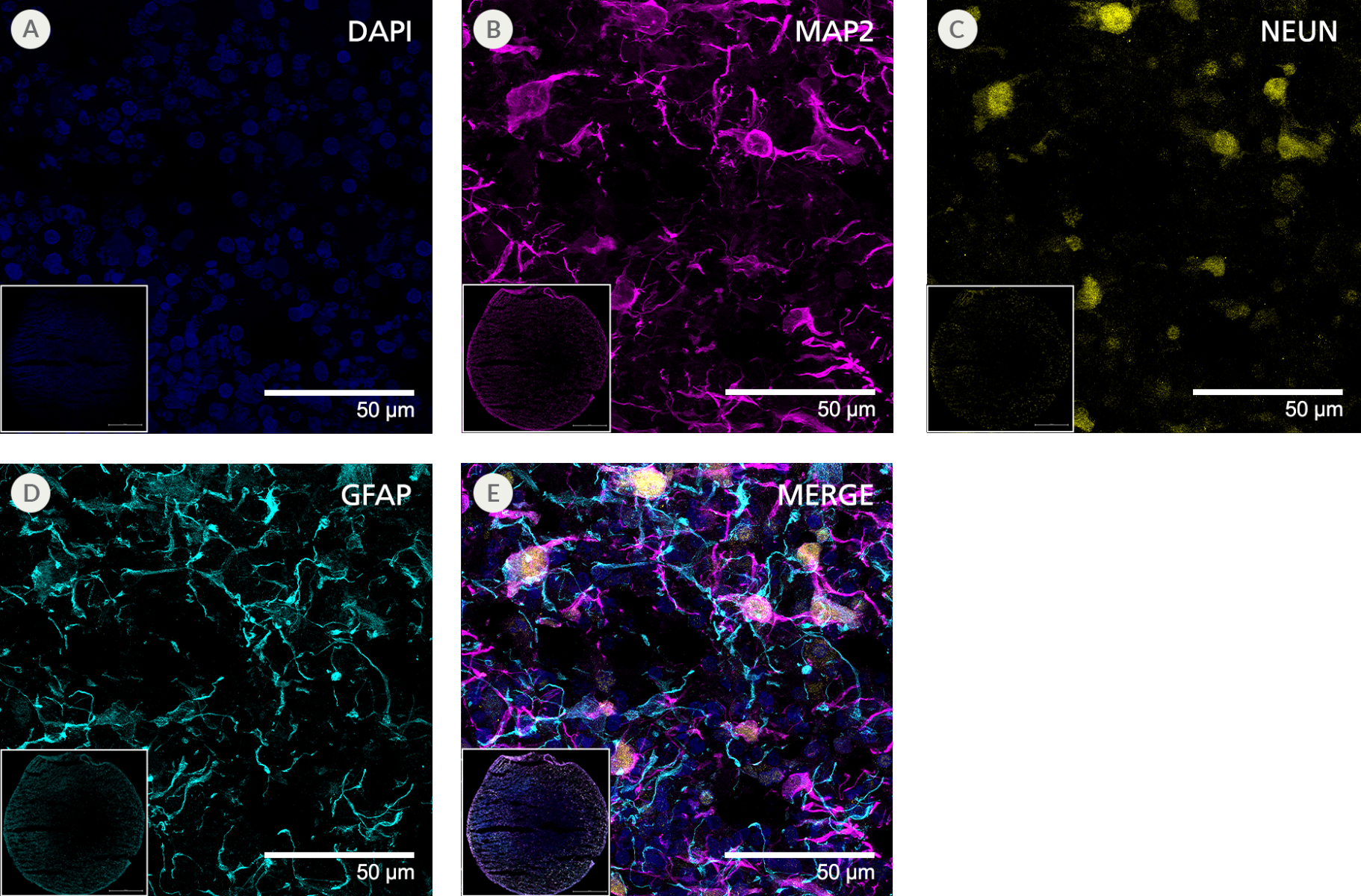
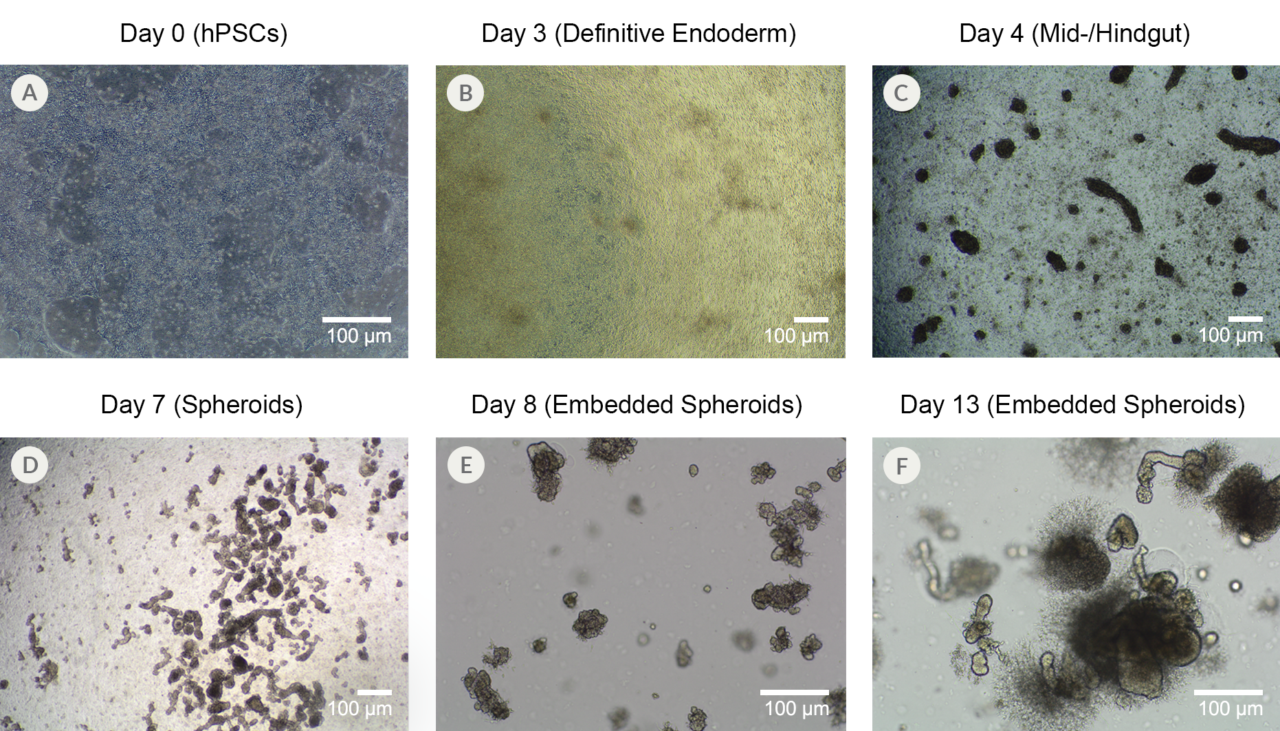
SCTi003-A细胞系已验证可在PBS-MINI生物反应器中扩增,为您培养物的高效放大提供了一条途径。该细胞系也已在二维和类器官模型中,使用STEMdiff™试剂盒在多种谱系和组织类型中进行了验证(图6-11)。浏览TeSR™和STEMdiff™细胞培养基产品,为您的细胞培养系统建立完整的工作流程。有关iPSC对照系的其他基因型,请查看我们所有的优质、健康的对照人iPSC系(女性和男性)。
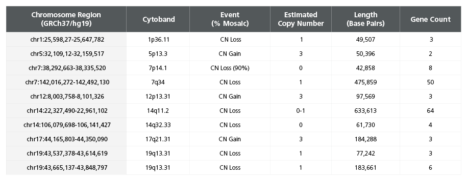
本产品仅供研究使用 (RUO),已获准用于学术和商业用途。血液样本的采集符合伦理规范,并已获得机构审查委员会 (IRB) 或其他监管机构批准的知情同意书和方案。有关供体详细信息和源细胞库的细胞质量鉴定,请参阅此页面上的数据图表。SCTi003-A源自αβ T细胞,并已进行 VDJ 序列重排。有关更多详细信息,请参阅批次特异性的分析证书以及有关诱导多能干细胞系的常见问题解答。
可根据要求提供全外显子组和全基因组序列数据文件。请联系我们获取报价。
此外,我们为SCTi003-A提供CRISPR-Cas9基因组编辑服务,以支持您的自定义模型开发。请填写此表格以讨论您的自定义基因组编辑项目要求。
某些产品仅在特定国家/地区提供。请联系您当地的销售代表或技术支持部门(邮箱:techsupport@stemcell.com)以获取更多信息。
包含
CryoStor® CS10
分类
冻存
细胞类型
多能干细胞
种属
人
细胞和组织来源
外周血,多能干细胞
品牌
STEMdiff,TeSR
研究领域
细胞系制备,疾病建模,药物发现和毒理检测,感染性疾病(传染病),神经科学,类器官,干细胞生物学
供体状态
正常
纯度
流式细胞术检测: TRA-1-60+ 和 OCT4+ ≥ 80%
请在《产品说明书》中查找相关支持信息和使用说明,或浏览下方更多实验方案。
本产品专为以下研究领域设计,适用于工作流程中的高亮阶段。探索这些工作流程,了解更多我们为各研究领域提供的其他配套产品。
| 物种 | 人类 |
|---|---|
| Contains | CryoStor® CS10 |
| 纯度 | ≥ 80% TRA-1-60+ and OCT4+ by flow cytometry |
| 细胞与组织来源 | 外周血, 多能干细胞 |
| 捐献者身份 | 正常 |
cGMP级、无酶的人多能干细胞选择与传代试剂
cGMP,稳定无饲养层维持培养基,适用于人ES和iPS 细胞
全自动细胞解冻系统,确保一致的解冻性能
<p>用于提高人胚胎干细胞和诱导多能干细胞在单细胞工作流程中存活率的添加物</p>
法律声明:
LIMITED USE LICENSEThese hiPSCs and their modifications (including but not limited to derivatives or differentiated progeny) shall not be used or administered in (1) human subjects for human clinical use; (2) animals for veterinary use for therapeutic, diagnostic, or prophylactic purposes or (3) any subject in relation to clinical applications, cell therapy, transplantation, and/or regenerative medicines, without limiting the generality of the foregoing.These hiPSCs and their modifications (including but not limited to derivatives or differentiated progeny) may not be used for monetization or commercialization purposes, including without limitation, used to, or with the goal to, perform services or supply products or rights, including in the manufacture of cellular therapies or other therapeutics, for monetary gain or the generation of royalties, revenues, sales or other valuable consideration. For clarity, these hiPSCs and their modifications (including but not limited to derivatives or differentiated progeny) may not be used for screening compounds, antibodies, proteins or peptides, except for the purposes of target discovery, target validation, or assay development, provided such activities and the results of such activities are not further used for monetization or commercialization purposes. It may be possible to obtain a further license for the prohibited uses referred to in this Limited Use License. Please contact iPSCrequests@stemcell.com for more details.
质量声明:
产品仅供研究使用,不用于针对人或动物的诊断或治疗。
在线联系


 沪公网安备31010102008431号