F. Cadamuro et al. (Feb 2023)
Carbohydrate polymers 302 120395
3D bioprinted colorectal cancer models based on hyaluronic acid and signalling glycans.
In cancer microenvironment,aberrant glycosylation events of ECM proteins and cell surface receptors occur. We developed a protocol to generate 3D bioprinted models of colorectal cancer (CRC) crosslinking hyaluronic acid and gelatin functionalized with three signalling glycans characterized in CRC,3'-Sialylgalactose,6'-Sialylgalactose and 2'-Fucosylgalactose. The crosslinking,performed exploiting azide functionalized gelatin and hyaluronic acid and 4arm-PEG-dibenzocyclooctyne,resulted in biocompatible hydrogels that were 3D bioprinted with commercial CRC cells HT-29 and patient derived CRC tumoroids. The glycosylated hydrogels showed good 3D printability,biocompatibility and stability over the time. SEM and synchrotron radiation SAXS/WAXS analysis revealed the influence of glycosylation in the construct morphology,whereas MALDI-MS imaging showed that protein profiles of tumoroid cells vary with glycosylation,indicating that sialylation and fucosylation of ECM proteins induce diverse alterations to the proteome of the tumoroid and surrounding cells.
View Publication
Brandl M et al. (AUG 1999)
Experimental hematology 27 8 1264--70
Bispecific antibody fragments with CD20 X CD28 specificity allow effective autologous and allogeneic T-cell activation against malignant cells in peripheral blood and bone marrow cultures from patients with B-cell lineage leukemia and lymphoma.
Bispecific antibodies directed against tumor-associated target antigens and to surface receptors mediating T-cell activation,such as the TCR/CD3 complex and the costimulatory receptor CD28,are capable of mediating T-cell activation resulting in tumor cell killing. In this study,we used the B-cell-associated antigens CD19 and CD20 as target structures on human leukemic cells. We found that a combination of bispecific antibody fragments (bsFab2) with target x CD3 and target x CD28 specificity induces vigorous autologous T-cell activation and killing of malignant cells in peripheral blood and bone marrow cultures from patients with chronic lymphocytic leukemia and follicular lymphoma. The bsFab2 targeting CD20 were considerably more effective than those binding to CD19. The colony-forming capacity of treated bone marrow was impaired due to large amounts of tumor necrosis factor alpha produced during bsFab2-induced T-cell activation. Neutralizing tumor necrosis factor alpha antibodies were found to reverse this negative effect without affecting T-cell activation and tumor cell killing. CD20 x CD28 bsFab2,when used alone rather than in combination,markedly improved the recognition of leukemic cells by allogeneic T cells. Therefore,these reagents may be capable of enhancing the immunogenicity of leukemic cells in general and,in particular,of increasing the antileukemic activity of allogeneic donor buffy coat cells in relapsed bone marrow transplanted patients.
View Publication
产品号#:
04431
产品名:
MethoCult™ H4431
Kumagai T et al. (JUN 2003)
Journal of the National Cancer Institute 95 12 896--905
Vitamin D2 analog 19-nor-1,25-dihydroxyvitamin D2: antitumor activity against leukemia, myeloma, and colon cancer cells.
BACKGROUND: 1,25-Dihydroxyvitamin D(3) inhibits growth of several types of human cancer cells in vitro,but its therapeutic use is hampered because it causes hypercalcemia. 19-nor-1,25-Dihydroxyvitamin D(2) (paricalcitol) is a noncalcemic vitamin D analog that is approved by the Food and Drug Administration for the treatment of secondary hyperparathyroidism. We investigated the antitumor activity and mechanism of action of paricalcitol in vitro and in vivo. METHODS: Effects of paricalcitol on proliferation,the cell cycle,differentiation,and apoptosis were examined in cancer cell lines. Effects on tumor growth were examined with colon cancer cell xenografts in nude mice (five in the experimental group and five in the control group). The interaction of paricalcitol with the vitamin D receptor (VDR) in mononuclear spleen cells and myeloid stem cells from wild-type and VDR knockout mice was examined. All statistical tests were two-sided. RESULTS: Paricalcitol inhibited the proliferation of myeloid leukemia cell lines HL-60,NB-4,and THP-1 cells at an effective dose that inhibited growth 50% (ED(50)) of 2.4-5.8 x 10(-9) M by inducing cell cycle arrest and differentiation. Paricalcitol inhibited the proliferation of NCI-H929 myeloma cells at an ED(50) of 2.0 x 10(-10) M by inducing cell cycle arrest and apoptosis. Paricalcitol also inhibited the proliferation of colon cancer cell lines HT-29 (ED(50) = 1.7 x 10(-8) M) and SW837 (ED(50) = 3.2 x 10(-8) M). HT-29 colon cancer xenografts in paricalcitol-treated nude mice were smaller (1044 mm(3) and 1752 mm(3),difference = 708 mm(3),95% confidence interval = 311 to 1104 mm(3); P =.03) and weighed less (1487 mg and 4162 mg,difference = 2675 mg,95% confidence interval = 2103 to 3248 mg; Ptextless.001) than those in vehicle-treated mice. Paricalcitol induced committed myeloid hematopoietic stem cells from wild-type but not from VDR knockout mice to differentiate as macrophages. CONCLUSION: Paricalcitol has anticancer activity against myeloid leukemia,myeloma,and colon cancer cells that may be mediated through the VDR. Because it has been approved by the Food and Drug Administration,clinical trials of this agent in certain cancers are reasonable.
View Publication

 EasySep™小鼠TIL(CD45)正选试剂盒


 挂图Small Molecules, Big Impact in Pluripotent Stem Cell Research Overview of signaling pathways and small molecules in pluripotent stem cell research
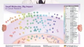 挂图Small Molecules, Big Impact in Cancer Research Overview of signaling pathways and small molecules in cancer research
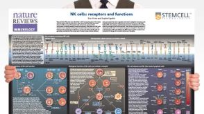 挂图Natural Killer Cells Overview of NK cell receptors, subsets, activation and function

 沪公网安备31010102008431号