Rega A et al. (MAR 2013)
Journal of immunology (Baltimore,Md. : 1950) 190 5 2391--402
Plasmacytoid dendritic cells play a key role in tumor progression in lipopolysaccharide-stimulated lung tumor-bearing mice.
The antitumor activity of LPS was first described by Dr. William Coley. However,its role in lung cancer remains unclear. The aim of our study was to elucidate the dose-dependent effects of LPS (0.1-10 μg/mouse) in a mouse model of B16-F10-induced metastatic lung cancer. Lung tumor growth increased at 3 and 7 d after the administration of low-dose LPS (0.1 μg/mouse) compared with control mice. This was associated with an influx of plasmacytoid dendritic cells (pDCs),regulatory T cells,myeloid-derived suppressor cells,and CD8(+) regulatory T cells. In contrast,high-dose LPS (10 μg/mouse) reduced lung tumor burden and was associated with a greater influx of pDCs,as well as a stronger Th1 and Th17 polarization. Depletion of pDCs during low-dose LPS administration resulted in a decreased lung tumor burden. Depletion of pDCs during high-dose LPS treatment resulted in an increased tumor burden. The dichotomy in LPS effects was due to the phenotype of pDCs,which were immunosuppressive after the low-dose LPS,and Th1- and T cytotoxic-polarizing cells after the high-dose LPS. Adoptive transfer of T cells into nude mice demonstrated that CD8(+) T cells were responsible for pDC recruitment following low-dose LPS administration,whereas CD4(+) T cells were required for pDC influx after the high-dose LPS. In conclusion,our data suggest differential effects of low-dose versus high-dose LPS on pDC phenotype and tumor progression or regression in the lungs of mice.
View Publication
产品号#:
19752
19752RF
19753
19753RF
19764
19764RF
产品名:
EasySep™小鼠浆细胞样DC分选试剂盒
RoboSep™ 小鼠浆细胞样DC分选试剂盒
Dannull J et al. (JUL 2013)
The Journal of clinical investigation 123 7 3135--45
Melanoma immunotherapy using mature DCs expressing the constitutive proteasome.
BACKGROUND Many cancers,including melanoma,exclusively express constitutive proteasomes (cPs) and are unable to express immunoproteasomes (iPs). In contrast,mature DCs used for immunotherapy exclusively express iPs. Since proteasomes generate peptides presented by HLA class I molecules,we hypothesized that mature melanoma antigen-loaded DCs engineered to process antigens through cPs would be superior inducers of antimelanoma immunity in vivo. METHODS Subjects with metastatic melanoma were vaccinated with mature DCs transfected with RNAs encoding melanoma antigens MART1,MAGE-3,gp100,and tyrosinase. These DCs were derived from monocytes that were untransfected (Arm A; n = 4),transfected with control siRNA (Arm B; n = 3),or transfected with siRNAs targeting the 3 inducible iP subunits (Arm C; n = 5). RESULTS Vaccination stimulated antigen-specific T cell responses in all subjects,which peaked after 3-4 vaccinations,but remained elevated in Arm C subjects. Also in Arm C,circulating melanoma cell levels (as detected by quantitative PCR) fell,and T cell lytic activity against autologous melanoma was induced. In HLA-A2 subjects,CD8 T cells that bound tetramers loaded with cP-derived melanoma antigenic peptides were found in the peripheral blood only in Arm C subjects. Of 2 subjects with active disease (both in Arm C),one had a partial clinical response,while the other,who exhibited diffuse dermal and soft tissue metastases,had a complete response. CONCLUSION These results suggest that the efficacy of melanoma DC-based immunotherapy is enhanced when tumor antigen-loaded DCs used for vaccination express cPs. TRIAL REGISTRATION Clinicaltrials.gov NCT00672542. FUNDING Duke Clinical Research Institute/Duke Translational Medicine Institute,Duke Melanoma Consortium,and Duke University Department of Surgery.
View Publication
Podrazil M et al. (JUL 2015)
Oncotarget 6 20 18192--205
Phase I/II clinical trial of dendritic-cell based immunotherapy (DCVAC/PCa) combined with chemotherapy in patients with metastatic, castration-resistant prostate cancer.
PURPOSE We conducted an open-label,single-arm Phase I/II clinical trial in metastatic CRPC (mCRPC) patients eligible for docetaxel combined with treatment with autologous mature dendritic cells (DCs) pulsed with killed LNCaP prostate cancer cells (DCVAC/PCa). The primary and secondary endpoints were safety and immune responses,respectively. Overall survival (OS),followed as a part of the safety evaluation,was compared to the predicted OS according to the Halabi and MSKCC nomograms. EXPERIMENTAL DESIGN Twenty-five patients with progressive mCRPC were enrolled. Treatment comprised of initial 7 days administration of metronomic cyclophosphamide 50 mg p.o. DCVAC/PCa treatment consisted of a median twelve doses of 1 × 10⁷ dendritic cells per dose injected s.c. (Aldara creme was applied at the site of injection) during a one-year period. The initial 2 doses of DCVAC/PCa were administered at a 2-week interval,followed by the administration of docetaxel (75 mg/m2) and prednisone (5 mg twice daily) given every 3 weeks until toxicity or intolerance was observed. The DCVAC/PCa was then injected every 6 weeks up to the maximum number of doses manufactured from one leukapheresis. RESULTS No serious DCVAC/PCa-related adverse events have been reported. The median OS was 19 months,whereas the predicted median OS was 11.8 months with the Halabi nomogram and 13 months with the MSKCC nomogram. Kaplan-Meier analyses showed that patients had a lower risk of death compared with both MSKCC (Hazard Ratio 0.26,95% CI: 0.13-0.51) and Halabi (Hazard Ratio 0.33,95% CI: 0.17-0.63) predictions. We observed a significant decrease in Tregs in the peripheral blood. The long-term administration of DCVAC/PCa led to the induction and maintenance of PSA specific T cells. We did not identify any immunological parameter that significantly correlated with better OS. CONCLUSIONS In patients with mCRPC,the combined chemoimmunotherapy with DCVAC/PCa and docetaxel was safe and resulted in longer than expected survival. Concomitant chemotherapy did not preclude the induction of specific anti-tumor cytotoxic T cells.
View Publication

 EasySep™小鼠TIL(CD45)正选试剂盒


 产品手册Highway1™: Fast, Gentle, and Automated Cell Sorting for Every Lab
 技术公告Achieve Scalable, High-Quality Nucleic Acid Extractions with the EasySep™ Total Nucleic Acid Extraction Kit
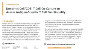 技术公告Dendritic Cell/CD8+ T Cell Co-Culture to Assess Antigen-Specific T Cell Functionality
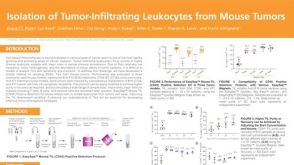 科学海报Isolation of Tumor-Infiltrating Leukocytes from Mouse Tumors


 沪公网安备31010102008431号