
 EasySep™小鼠TIL(CD45)正选试剂盒


技术资料
-
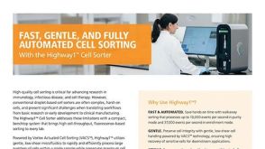 产品手册Highway1™: Fast, Gentle, and Automated Cell Sorting for Every Lab
品牌:
Highway1
发布日期: 04/14/2026 -
-
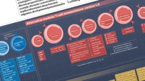 挂图T Cell Nomenclature: From Subsets to Modules A modular framework for classifying T cells by lineage, function, migration, differentiation, and antigen context.发布日期: 12/05/2025
 -
 研究综述The Predictive Power of Organoid-Based New Approach Methodologies in Drug Discovery
细胞类型:
上皮细胞,多能干细胞,肠道细胞,肾细胞,胰腺细胞,PSC衍生上皮细胞,PSC衍生肝细胞,呼吸道细胞
发布日期: 10/31/2025 -
 技术公告Achieve Scalable, High-Quality Nucleic Acid Extractions with the EasySep™ Total Nucleic Acid Extraction Kit
细胞类型:
B细胞,NK细胞,T细胞,其他细胞系,单个核细胞,单核细胞,巨噬细胞,树突状细胞(DCs),淋巴细胞,癌细胞及细胞系,粒细胞及其亚群,肿瘤细胞
发布日期: 09/11/2025 -
-
 24:08
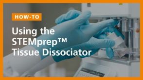
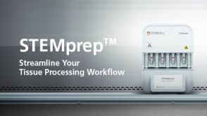
线上讲座Streamline Your Tissue Processing Using the Automated STEMprep™ Tissue Dissociator发布日期: 06/12/2025
24:08
线上讲座Streamline Your Tissue Processing Using the Automated STEMprep™ Tissue Dissociator发布日期: 06/12/2025 -
-
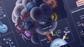 挂图Rational Combination of Cancer Therapies with PD1 Axis Blockade Lists the ways to combine new immunotherapeutic targets and modalities for your translational research.发布日期: 10/31/2024
 -

 沪公网安备31010102008431号