
 EasySep™小鼠TIL(CD45)正选试剂盒


技术资料
-

-
 1:07:14
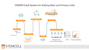
线上讲座Optimized Workflows for High-Efficiency Genome Editing in Stem and Primary Cell Types发布日期: 09/09/2019
1:07:14
线上讲座Optimized Workflows for High-Efficiency Genome Editing in Stem and Primary Cell Types发布日期: 09/09/2019 -
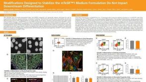 科学海报Modifications Designed to Stabilize the mTeSR1™ Formulation Do Not Impact Downstream Differentiation
Conference:
ISSCR 2019
发布日期: 08/09/2019 -
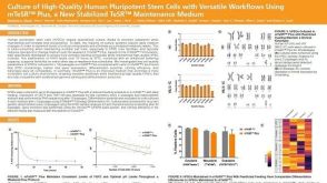 科学海报Culture of High-Quality Human Pluripotent Stem Cells with Versatile Workflows Using mTeSR™ Plus, a New Stabilized TeSR™ Maintenance Medium
Conference:
ISSCR 2019
发布日期: 07/23/2019 -
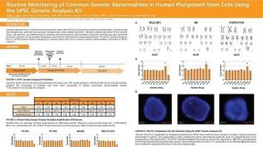 科学海报Routine Monitoring of Common Genetic Abnormalities in Human Pluripotent Stem Cells Using the hPSC Genetic Analysis Kit
Conference:
ISSCR 2019
发布日期: 07/23/2019 -
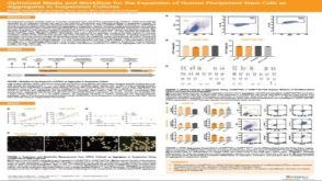 科学海报Optimized Media and Workflow for the Expansion of Human Pluripotent Stem Cells as Aggregates in Suspension Cultures
Conference:
ISSCR 2019
发布日期: 07/05/2019

 沪公网安备31010102008431号