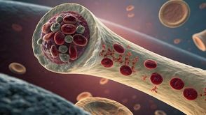
Hoxa3 promotes the differentiation of hematopoietic progenitor cells into proangiogenic Gr-1+CD11b+ myeloid cells.
Injury induces the recruitment of bone marrow-derived cells (BMDCs) that contribute to the repair and regeneration process. The behavior of BMDCs in injured tissue has a profound effect on repair,but the regulation of BMDC behavior is poorly understood. Aberrant recruitment/retention of these cells in wounds of diabetic patients and animal models is associated with chronic inflammation and impaired healing. BMD Gr-1(+)CD11b(+) cells function as immune suppressor cells and contribute significantly to tumor-induced neovascularization. Here we report that Gr-1(+)CD11b(+) cells also contribute to injury-induced neovascularization,but show altered recruitment/retention kinetics in the diabetic environment. Moreover,diabetic-derived Gr-1(+)CD11b(+) cells fail to stimulate neovascularization in vivo and have aberrant proliferative,chemotaxis,adhesion,and differentiation potential. Previously we demonstrated that gene transfer of HOXA3 to wounds of diabetic mice is taken up by and expressed by recruited BMDCs. This is associated with a suppressed inflammatory response,enhanced neovascularization,and accelerated wound healing. Here we show that sustained expression of Hoxa3 in diabetic-derived BMD Gr-1(+)CD11b(+) cells reverses their diabetic phenotype. These findings demonstrate that manipulation of adult stem/progenitor cells ex vivo could be used as a potential therapy in patients with impaired wound healing.
View Publication
Novel imatinib-sensitive PDGFRA-activating point mutations in hypereosinophilic syndrome induce growth factor independence and leukemia-like disease.
The FIP1L1-PDGFRA fusion is seen in a fraction of cases with a presumptive diagnosis of hypereosinophilic syndrome (HES). However,because most HES patients lack FIP1L1-PDGFRA,we studied whether they harbor activating mutations of the PDGFRA gene. Sequencing of 87 FIP1L1-PDGFRA-negative HES patients revealed several novel PDGFRA point mutations (R481G,L507P,I562M,H570R,H650Q,N659S,L705P,R748G,and Y849S). When cloned into 32D cells,N659S and Y849S and-on selection for high expressors-also H650Q and R748G mutants induced growth factor-independent proliferation,clonogenic growth,and constitutive phosphorylation of PDGFRA and Stat5. Imatinib antagonized Stat5 phosphorylation. Mutations involving positions 659 and 849 had been shown previously to possess transforming potential in gastrointestinal stromal tumors. Because H650Q and R748G mutants possessed only weak transforming activity,we injected 32D cells harboring these mutants or FIP1L1-PDGFRA into mice and found that they induced a leukemia-like disease. Oral imatinib treatment significantly decreased leukemic growth in vivo and prolonged survival. In conclusion,our data provide evidence that imatinib-sensitive PDGFRA point mutations play an important role in the pathogenesis of HES and we propose that more research should be performed to further define the frequency and treatment response of PDGFRA mutations in FIP1L1-PDGFRA-negative HES patients.
View Publication
产品号#:
03231
产品名:
MethoCult™ M3231
N'jai AU et al. (APR 2011)
Molecular pharmacology 79 4 724--34
Acute disruption of bone marrow hematopoiesis by benzo(a)pyrene is selectively reversed by aryl hydrocarbon receptor-mediated processes.
Bone marrow (BM) hematopoietic cells are selectively sensitive to polycyclic aromatic hydrocarbons (PAH) in vivo. 7,12-Dimethylbenz(a)anthracene (DMBA),but not benzo(a)pyrene (BP),depletes BM hematopoietic cells in C57BL/6 mice. This difference is due to a BP-selective aryl hydrocarbon receptor (AhR)-mediated recovery. Colony-forming unit assays show suppression of lymphoid progenitors by each PAH within 6 h but a subsequent recovery,exclusively after BP treatment. Suppression of myeloid progenitors (6 h) occurs only for DMBA. Each progenitor responded equally to DMBA and BP in congenic mice expressing the PAH-resistant AhR (AhR(d)). AhR,therefore,mediates this BP recovery in each progenitor type. These PAH suppressions depend on Cyp1b1-mediated metabolism. Paradoxically,few genes responded to DMBA,whereas 12 times more responded to BP. Progenitor suppression by DMBA,therefore,occurs with minimal effects on the general BM population. Standard AhR-mediated stimulations (Cyp1a1,Cyp1b1,Ahrr) were similar for each PAH and for the specific agonist 2,3,7,8-tetrachlorodibenzo-p-dioxin but were absent in AhR(d) mice. A group of 12 such AhR responses was sustained from 6 to 24 h. A second,larger set of BP responses (chemokines,cytokines,cyclooxygenase 2) differed in two respects; DMBA responses were low and BP responses declined extensively from 6 to 24 h. A third cluster exhibited BP-induced increases in protective genes (Nqo1,GST-mu) that appeared only after 12 h. Conversion of BP to quinones contributes oxidative signaling not seen with DMBA. We propose that genes in this second cluster,which share oxidative signaling and AhR activation,provide the AhR-dependent protection of hematopoietic progenitors seen for BP.
View Publication
产品号#:
03534
03630
产品名:
MethoCult™ GF M3534
MethoCult™ M3630
Kim M-H et al. (MAR 2011)
Blood 117 12 3343--52
Neutrophil survival and c-kit(+)-progenitor proliferation in Staphylococcus aureus-infected skin wounds promote resolution.
Polymorphonuclear neutrophils (PMNs) are critical for the formation,maintenance,and resolution of bacterial abscesses. However,the mechanisms that regulate PMN survival and proliferation during the evolution of an abscess are not well defined. Using a mouse model of Staphylococcus aureus abscess formation within a cutaneous wound,combined with real-time imaging of genetically tagged PMNs,we observed that a high bacterial burden elicited a sustained mobilization of PMNs from the bone marrow to the infected wound,where their lifespan was markedly extended. A continuous rise in wound PMN number,which was not accounted for by trafficking from the bone marrow or by prolonged survival,was correlated with the homing of c-kit(+)-progenitor cells from the blood to the wound,where they proliferated and formed mature PMNs. Furthermore,by blocking their recruitment with an antibody to c-kit,which severely limited the proliferation of mature PMNs in the wound and shortened mouse survival,we confirmed that progenitor cells are not only important contributors to PMN expansion in the wound,but are also functionally important for immune protection. We conclude that the abscess environment provides a niche capable of regulating PMN survival and local proliferation of bone marrow-derived c-kit(+)-progenitor cells.
View Publication
产品号#:
03434
03444
产品名:
MethoCult™ GF M3434
MethoCult™ GF M3434
Yoshimi A et al. (MAR 2011)
Blood 117 13 3617--28
Evi1 represses PTEN expression and activates PI3K/AKT/mTOR via interactions with polycomb proteins.
Evi1 (ecotropic viral integration site 1) is essential for proliferation of hematopoietic stem cells and implicated in the development of myeloid disorders. Particularly,high Evi1 expression defines one of the largest clusters in acute myeloid leukemia and is significantly associated with extremely poor prognosis. However,mechanistic basis of Evi1-mediated leukemogenesis has not been fully elucidated. Here,we show that Evi1 directly represses phosphatase and tensin homologue deleted on chromosome 10 (PTEN) transcription in the murine bone marrow,which leads to activation of AKT/mammalian target of rapamycin (mTOR) signaling. In a murine bone marrow transplantation model,Evi1 leukemia showed modestly increased sensitivity to an mTOR inhibitor rapamycin. Furthermore,we found that Evi1 binds to several polycomb group proteins and recruits polycomb repressive complexes for PTEN down-regulation,which shows a novel epigenetic mechanism of AKT/mTOR activation in leukemia. Expression analyses and ChIPassays with human samples indicate that our findings in mice models are recapitulated in human leukemic cells. Dependence of Evi1-expressing leukemic cells on AKT/mTOR signaling provides the first example of targeted therapeutic modalities that suppress the leukemogenic activity of Evi1. The PTEN/AKT/mTOR signaling pathway and the Evi1-polycomb interaction can be promising therapeutic targets for leukemia with activated Evi1.
View Publication

 EasySep™小鼠TIL(CD45)正选试剂盒


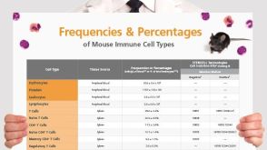 挂图Frequencies and Percentages of Mouse Immune Cell Types List of the frequencies of over 25 immune cell types in C57BL/6 mice
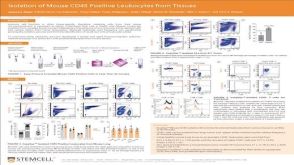


 沪公网安备31010102008431号