
 EasySep™小鼠TIL(CD45)正选试剂盒


技术资料
-
-
-
-
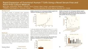 科学海报Rapid Expansion of Functional Human T Cells Using a Novel Serum-Free and Xeno-Free Culture Medium
Conference:
CCIC 2015
-
-
 科学海报A Specialized Tube to Make Enrichment of Specific Cell Subsets Faster and Easier
Conference:
BSHI 2012,EFI 2012,IHIW 2012,AAI 2012
-
 科学海报One-Step Enrichment of Leukocyte Subsets Directly in the Blood Collection Tube
Conference:
AAI 2003
-
 科学海报Column-Free Isolation of Highly Purified and Functional Human Regulatory T Cells
Conference:
ASI 2010
-
 科学海报Isolation of Highly Purified Mouse CD4+CD25+Foxp3+ Regulatory T Cells in Less
Conference:
AAI 2014
-
 科学海报Immunomagnetic Isolation Method for Mouse CD4+CD25+ Regulatory T Cells
Conference:
AAI 2008,CSI 2008
-
-

 沪公网安备31010102008431号