M. Riopel et al. ( 2019)
Molecular metabolism 20 89--101
CX3CL1-Fc treatment prevents atherosclerosis in Ldlr KO mice.
OBJECTIVE Atherosclerosis is a major cause of cardiovascular disease. Monocyte-endothelial cell interactions are partly mediated by expression of monocyte CX3CR1 and endothelial cell fractalkine (CX3CL1). Interrupting the interaction between this ligand-receptor pair should reduce monocyte binding to the endothelial wall and reduce atherosclerosis. We sought to reduce atherosclerosis by preventing monocyte-endothelial cell interactions through use of a long-acting CX3CR1 agonist. METHODS In this study,the chemokine domain of CX3CL1 was fused to the mouse Fc region to generate a long-acting soluble form of CX3CL1 suitable for chronic studies. CX3CL1-Fc or saline was injected twice a week (30 mg/kg) for 4 months into Ldlr knockout (KO) mice on an atherogenic western diet. RESULTS CX3CL1-Fc-treated Ldlr KO mice showed decreased en face aortic lesion surface area and reduced aortic root lesion size with decreased necrotic core area. Flow cytometry analyses of CX3CL1-Fc-treated aortic wall cell digests revealed a decrease in M1-like polarized macrophages and T cells. Moreover,CX3CL1-Fc administration reduced diet-induced atherosclerosis after switching from an atherogenic to a normal chow diet. In vitro monocyte adhesion studies revealed that CX3CL1-Fc treatment caused fewer monocytes to adhere to a human umbilical vein endothelial cell monolayer. Furthermore,a dorsal window chamber model demonstrated that CX3CL1-Fc treatment decreased in vivo leukocyte adhesion and rolling in live capillaries after short-term ischemia-reperfusion. CONCLUSION These results indicate that CX3CL1-Fc can inhibit monocyte/endothelial cell adhesion as well as reduce atherosclerosis.
View Publication
文献
N. Kuroda et al. (jun 2019)
Scientific reports 9 1 8568
Infiltrating CCR2+ monocytes and their progenies, fibrocytes, contribute to colon fibrosis by inhibiting collagen degradation through the production of TIMP-1.
Intestinal fibrosis is a serious complication in inflammatory bowel disease (IBD). Despite the remarkable success of recent anti-inflammatory therapies for IBD,incidence of intestinal fibrosis and need for bowel resection have not significantly changed. To clarify the contribution of haematopoietic-derived cells in intestinal fibrosis,we prepared bone marrow (BM) chimeric mice (chimeras),which were reconstituted with BM cells derived from enhanced green fluorescent protein (EGFP)-transgenic mice or CC chemokine receptor 2 (CCR2)-deficient mice. After 2 months of transplantation,BM chimeras were treated with azoxymethane/dextran sodium sulphate. During chronic inflammation,CCR2+ BM-derived monocyte and fibrocyte infiltration into the colon and CC chemokine ligand 2 production increased,leading to colon fibrosis in EGFP BM chimeras. In CCR2-deficient BM chimeras,monocyte and fibrocyte numbers in the colonic lamina propria significantly decreased,and colon fibrosis was attenuated. In colon tissue,mRNA expression of tissue inhibitor of metalloproteinase (TIMP)-1 but not of collagen I,transforming growth factor-beta1 or matrix metalloproteinases was significantly different between the two chimeras. CCR2+ monocytes and fibrocytes showed high Timp1 mRNA expression. Our results suggest that infiltrating CCR2+ monocytes and their progenies,fibrocytes,promote colon fibrosis by inhibiting collagen degradation through TIMP-1 production.
View Publication
文献
D. Birkl et al. (jul 2019)
Mucosal immunology 12 4 909--918
TNFalpha promotes mucosal wound repair through enhanced platelet activating factor receptor signaling in the epithelium.
Pathobiology of several chronic inflammatory disorders,including ulcerative colitis and Crohn's disease is related to intermittent,spontaneous injury/ulceration of mucosal surfaces. Disease morbidity has been associated with pathologic release of the pro-inflammatory cytokine tumor necrosis factor alpha (TNFalpha). In this report,we show that TNFalpha promotes intestinal mucosal repair through upregulation of the GPCR platelet activating factor receptor (PAFR) in the intestinal epithelium. Platelet activating factor (PAF) was increased in healing mucosal wounds and its engagement with epithelial PAFR leads to activation of epidermal growth factor receptor,Src and Rac1 signaling to promote wound closure. Consistent with these findings,delayed colonic mucosal repair was observed after administration of a neutralizing TNFalpha antibody and in mice lacking PAFR. These findings suggest that in the injured mucosa,the pro-inflammatory milieu containing TNFalpha and PAF sets the stage for reparative events mediated by PAFR signaling.
View Publication
文献
S. Bhatia et al. (may 2019)
Cancer research 79 10 2722--2735
Inhibition of EphB4-Ephrin-B2 Signaling Reprograms the Tumor Immune Microenvironment in Head and Neck Cancers.
Identifying targets present in the tumor microenvironment that contribute to immune evasion has become an important area of research. In this study,we identified EphB4-ephrin-B2 signaling as a regulator of both innate and adaptive components of the immune system. EphB4 belongs to receptor tyrosine kinase family that interacts with ephrin-B2 ligand at sites of cell-cell contact,resulting in bidirectional signaling. We found that EphB4-ephrin-B2 inhibition alone or in combination with radiation (RT) reduced intratumoral regulatory T cells (Tregs) and increased activation of both CD8+ and CD4+Foxp3- T cells compared with the control group in an orthotopic head and neck squamous cell carcinoma (HNSCC) model. We also compared the effect of EphB4-ephrin-B2 inhibition combined with RT with combined anti-PDL1 and RT and observed similar tumor growth suppression,particularly at early time-points. A patient-derived xenograft model showed reduction of tumor-associated M2 macrophages and favored polarization towards an antitumoral M1 phenotype following EphB4-ephrin-B2 inhibition with RT. In vitro,EphB4 signaling inhibition decreased Ki67-expressing Tregs and Treg activation compared with the control group. Overall,our study is the first to implicate the role of EphB4-ephrin-B2 in tumor immune response. Moreover,our findings suggest that EphB4-ephrin-B2 inhibition combined with RT represents a potential alternative for patients with HNSCC and could be particularly beneficial for patients who are ineligible to receive or cannot tolerate anti-PDL1 therapy. SIGNIFICANCE: These findings present EphB4-ephrin-B2 inhibition as an alternative to anti-PDL1 therapeutics that can be used in combination with radiation to induce an effective antitumor immune response in patients with HNSCC.
View Publication
C. Petes et al. (SEP 2018)
Scientific Reports 8 1 13704
IL-27 amplifies cytokine responses to Gram-negative bacterial products and Salmonella typhimurium infection.
Cytokine responses from monocytes and macrophages exposed to bacteria are of particular importance in innate immunity. Focusing on the impact of the immunoregulatory cytokine interleukin (IL)-27 on control of innate immune system responses,we examined human immune responses to bacterial products and bacterial infection by E. coli and S. typhimurium. Since the effect of IL-27 treatment in human myeloid cells infected with bacteria is understudied,we treated human monocytes and macrophages with IL-27 and either LPS,flagellin,or bacteria,to investigate the effect on inflammatory signaling and cytokine responses. We determined that simultaneous stimulation with IL-27 and LPS derived from E. coli or S. typhimurium resulted in enhanced IL-12p40,TNF-$\alpha$,and IL-6 expression compared to that by LPS alone. To elucidate if IL-27 manipulated the cellular response to infection with bacteria,we infected IL-27 treated human macrophages with S. typhimurium. While IL-27 did not affect susceptibility to S. typhimurium infection or S. typhimurium-induced cell death,IL-27 significantly enhanced proinflammatory cytokine production in infected cells. Taken together,we highlight a role for IL-27 in modulating innate immune responses to bacterial infection.
View Publication
Freeman SA et al. (JAN 2018)
Cell 172 2-Jan 305--317.e10
Transmembrane Pickets Connect Cyto- and Pericellular Skeletons Forming Barriers to Receptor Engagement.
Phagocytic receptors must diffuse laterally to become activated upon clustering by multivalent targets. Receptor diffusion,however,can be obstructed by transmembrane proteins (pickets") that are immobilized by interacting with the cortical cytoskeleton. The molecular identity of these pickets and their role in phagocytosis have not been defined. We used single-molecule tracking to study the interaction between Fcγ receptors and CD44 an abundant transmembrane protein capable of indirect association with F-actin hence likely to serve as a picket. CD44 tethers reversibly to formin-induced actin filaments curtailing receptor diffusion. Such linear filaments predominate in the trailing end of polarized macrophages where receptor mobility was minimal. Conversely receptors were most mobile at the leading edge where Arp2/3-driven actin branching predominates. CD44 binds hyaluronan anchoring a pericellular coat that also limits receptor displacement and obstructs access to phagocytic targets. Force must be applied to traverse the pericellular barrier enabling receptors to engage their targets.
View Publication
文献
Dewhurst JA et al. (AUG 2017)
Scientific reports 7 1 7143
Characterisation of lung macrophage subpopulations in COPD patients and controls.
Lung macrophage subpopulations have been identified based on size. We investigated characteristics of small and large macrophages in the alveolar spaces and lung interstitium of COPD patients and controls. Alveolar and interstitial cells were isolated from lung resection tissue from 88 patients. Macrophage subpopulation cell-surface expression of immunological markers and phagocytic ability were assessed by flow cytometry. Inflammatory related gene expression was measured. Alveolar and interstitial macrophages had subpopulations of small and large macrophages based on size and granularity. Alveolar macrophages had similar numbers of small and large cells; interstitial macrophages were mainly small. Small macrophages expressed significantly higher cell surface HLA-DR,CD14,CD38 and CD36 and lower CD206 compared to large macrophages. Large alveolar macrophages showed lower marker expression in COPD current compared to ex-smokers. Small interstitial macrophages had the highest pro-inflammatory gene expression levels,while large alveolar macrophages had the lowest. Small alveolar macrophages had the highest phagocytic ability. Small alveolar macrophage CD206 expression was lower in COPD patients compared to smokers. COPD lung macrophages include distinct subpopulations; Small interstitial and small alveolar macrophages with more pro-inflammatory and phagocytic function respectively,and large alveolar macrophages with low pro-inflammatory and phagocytic ability.
View Publication
文献
Chen WLK et al. ( 2017)
Biotechnology and bioengineering 114 11 2648--2659
Integrated gut/liver microphysiological systems elucidates inflammatory inter-tissue crosstalk.
A capability for analyzing complex cellular communication among tissues is important in drug discovery and development,and in vitro technologies for doing so are required for human applications. A prominent instance is communication between the gut and the liver,whereby perturbations of one tissue can influence behavior of the other. Here,we present a study on human gut-liver tissue interactions under normal and inflammatory contexts,via an integrative multi-organ platform comprising human liver (hepatocytes and Kupffer cells),and intestinal (enterocytes,goblet cells,and dendritic cells) models. Our results demonstrated long-term (>2 weeks) maintenance of intestinal (e.g.,barrier integrity) and hepatic (e.g.,albumin) functions in baseline interaction. Gene expression data comparing liver in interaction with gut,versus isolation,revealed modulation of bile acid metabolism. Intestinal FGF19 secretion and associated inhibition of hepatic CYP7A1 expression provided evidence of physiologically relevant gut-liver crosstalk. Moreover,significant non-linear modulation of cytokine responses was observed under inflammatory gut-liver interaction; for example,production of CXCR3 ligands (CXCL9,10,11) was synergistically enhanced. RNA-seq analysis revealed significant upregulation of IFNα/β/γ signaling during inflammatory gut-liver crosstalk,with these pathways implicated in the synergistic CXCR3 chemokine production. Exacerbated inflammatory response in gut-liver interaction also negatively affected tissue-specific functions (e.g.,liver metabolism). These findings illustrate how an integrated multi-tissue platform can generate insights useful for understanding complex pathophysiological processes such as inflammatory organ crosstalk. Biotechnol. Bioeng. 2017;114: 2648-2659. textcopyright 2017 Wiley Periodicals,Inc.
View Publication

 EasySep™小鼠TIL(CD45)正选试剂盒


 文献
文献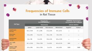 挂图Frequencies of Immune Cells in Rat Tissue Lists the estimated frequencies of more than 15 immune cell types in Sprague Dawley rats
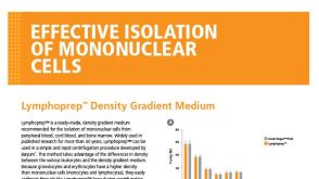 BrochureLymphoprep™ Density Gradient Medium for Mononuclear Cell Isolation

 沪公网安备31010102008431号