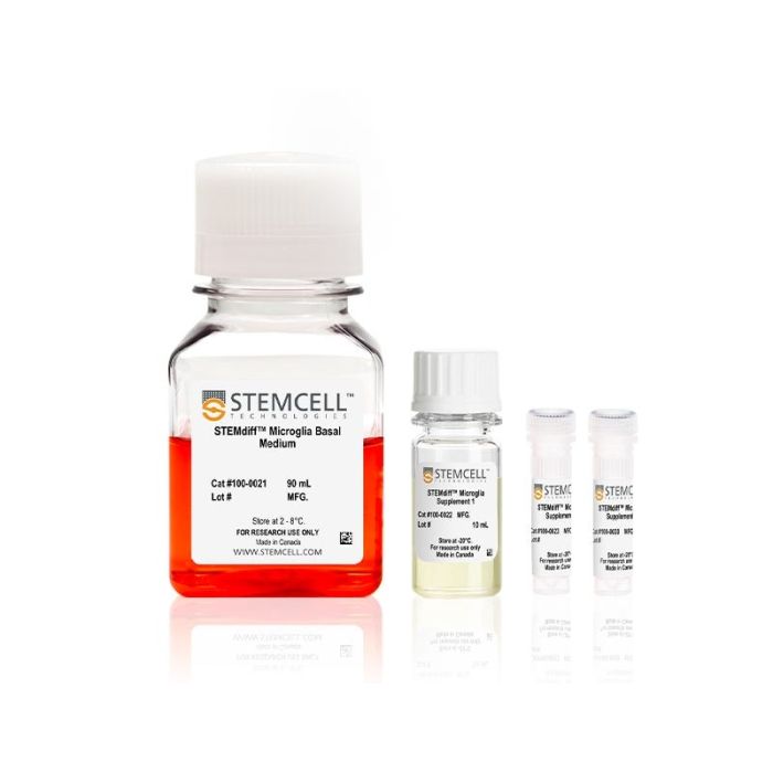
产品号 #100-0020_C
用于将人胚胎干细胞(ES)和诱导多能干细胞(iPS)来源的小胶质细胞前体分化成熟为小胶质细胞的成熟试剂盒。
若您需要咨询产品或有任何技术问题,请通过官方电话 400 885 9050 或邮箱 info.cn@stemcell.com 与我们联系。
用于将人胚胎干细胞(ES)和诱导多能干细胞(iPS)来源的小胶质细胞前体分化成熟为小胶质细胞的成熟试剂盒。
用于将人胚胎干细胞(ES)和诱导多能干细胞(iPS)来源的小胶质细胞前体分化成熟为小胶质细胞的成熟试剂盒。
STEMdiff™ 小胶质细胞培养系统包括 STEMdiff™ 小胶质细胞分化试剂盒和 STEMdiff™ 小胶质细胞成熟试剂盒。这两种试剂盒结合使用,用于分化和成熟采用 STEMdiff™ 造血试剂盒(货号 #05310)获得的人类多能干细胞 (hPSC) 衍生小胶质细胞。
该系统基于 Mathew Blurton-Jones 实验室的方案(Abud 等,2017),生成的细胞为高纯度的小胶质细胞群(>80% CD45/CD11b 阳性,>50% TREM2 阳性小胶质细胞;形态上明显不同的单核细胞或巨噬细胞占比 <20%)。
使用该系列产品获得的细胞可作为研究神经炎症、人类神经系统发育与疾病、共培养实验以及毒性测试的多功能工具。
分类
专用培养基
细胞类型
造血细胞,PSC衍生,小胶质细胞,神经细胞,PSC衍生
种属
人
应用
细胞培养,分化
品牌
STEMdiff
研究领域
疾病建模,药物发现和毒理检测,免疫,神经科学
制剂类别
无血清

Figure 1. Schematic for the STEMdiff™ Microglia Culture System Protocol
Microglial precursors can be generated in 24 days from hPSC-derived hematopoietic progenitor cells. For the generation of hematopoietic progenitor cells, see documentation for STEMdiff™ Hematopoietic Kit (Catalog #05310). For the maturation of microglial precursors to functional microglia, see the PIS.

Figure 2. Microglia Generated Using the STEMdiff™ Microglia Culture System Exhibit Robust Expansion, Mature Phenotypic Markers, and Homeostatic Morphology
(A) Microglia generated using the STEMdiff™ Microglia Culture System undergo a four-fold expansion, on average, across four cell lines. The fold expansion was calculated by taking the total cell count at Day 24 and dividing it by the number of seeded cells at Day 0. The bars show the mean ± standard deviation. Technical replicates were averaged, n = 1 - 4 technical replicates, 1 - 9 experimental setups.
(B) Microglia generated with STEMdiff™ Microglia Culture System have CD45+ CD11b+ co-expression and P2RY12+ expression as measured by flow cytometry on Day 24. The bars show the mean ± standard deviation.Technical replicates (n = 1 - 4) were averaged, and each dot in the graph represents an experimental replicate.
(C) Normal microglial morphology, characterized by small cell bodies and ramified processes, is observed in cells generated using the STEMdiff™ Microglia Culture System. Images at Days 12 and 24 were captured prior to replate and harvest. Scale bar = 100 µm.

Figure 3. Microglia Generated with STEMdiff™ Microglia Culture System Express Disease-Relevant Genes Similar to Those from Published Differentiation and Maturation Protocols
Bulk RNA-seq datasets were extracted from 8 different publications that generated hPSC- (iMGL) and primary- (MGL) derived microglia and their transcriptional profiles compared to data from microglia generated with STEMdiff™ Microglia Culture System. The heat map displays absolute expression levels for select genes associated with Alzheimer’s disease, Parkinson’s disease, and viral encephalitis. Significant differences in gene expression between microglia generated with STEMdiff™ Microglia Culture System and any of the other 3 groups were identified by differential gene expression analysis. *= p<0.05 (DEseq2, adjusted). hPSC = human pluripotent stem cell.

Figure 4. Microglia Generated with STEMdiff™ Microglia Culture System Release Cytokines in Response to Inflammatory Signals
Microglia were generated using the STEMdiff™ Microglia Culture System and stimulated with 100 ng/mL LPS for 24 hours. The release of pro-inflammatory (TNFα, IL-6, IFN-γ, IL-1β, GM-CSF, IL-12p70, IL-2, IL-8) and anti-inflammatory (IL-10) cytokines were measured by MSD. The microglia release cytokines in response to LPS treatment, as expected. *, p ≤ 0.05; **, p ≤ 0.01; ***, p ≤ 0.001; ****, p ≤ 0.0001. LPS = lipopolysaccharide; MSD = Meso Scale Discovery.

Figure 5. STEMdiff™ Microglia Culture System Generates Functional Microglia Capable of Phagocytosis at Day 34
Microglia taking up pH-sensitive bioindicator particles at a concentration of 5 μg/mL were measured over a 72-hour time period with live cell imaging. As the particles are phagocytosed, the particles turn red and are concentrated within the cells. Over time, the microglia display an activated ameboid morphology. Scale bar = 400 μm.

Figure 6. PSC-Derived Microglia Incorporate into Brain Organoids After 10 Days and Display an Activated Morphology upon Injury.
(A) Representative microglia and brain organoid co-cultures after 10 days, stained with IBA1 for microglia (green) and MAP2 for neurons (magenta). The microglia integrate among the neurons and display an unactivated morphology with extended processes (arrow).
(B) The microglia display an activated amoeboid morphology upon injury as shown by IBA1 staining.
请在《产品说明书》中查找相关支持信息和使用说明,或浏览下方更多实验方案。
本产品专为以下研究领域设计,适用于工作流程中的高亮阶段。探索这些工作流程,了解更多我们为各研究领域提供的其他配套产品。
Thank you for your interest in IntestiCult™ Organoid Growth Medium (Human). Please provide us with your contact information and your local representative will contact you with a customized quote. Where appropriate, they can also assist you with a(n):
Estimated delivery time for your area
Product sample or exclusive offer
In-lab demonstration
| 物种 | 人类 |
|---|---|
| 配方 | 无血清 |
无血清神经添加物(50X)
提升神经元功能的无血清基础培养基
用于人脑类器官建立与成熟的培养基试剂盒
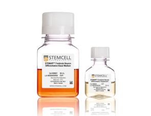
用于将人 ES 和 iPS 细胞衍生的神经前体细胞分化为神经元前体细胞的分化试剂盒
扫描二维码或搜索微信号STEMCELLTech,即可关注我们的微信平台,第一时间接收丰富的技术资源和最新的活动信息。
如您有任何问题,欢迎发消息给STEMCELLTech微信公众平台,或与我们通过电话/邮件联系:400 885 9050 INFO.CN@STEMCELL.COM。
在线联系

