
 EasySep™小鼠TIL(CD45)正选试剂盒


产品号 #100-0170_C
用于hPSC向NK细胞的扩增和分化
若您需要咨询产品或有任何技术问题,请通过官方电话 400 885 9050 或邮箱 info.cn@stemcell.com 与我们联系。
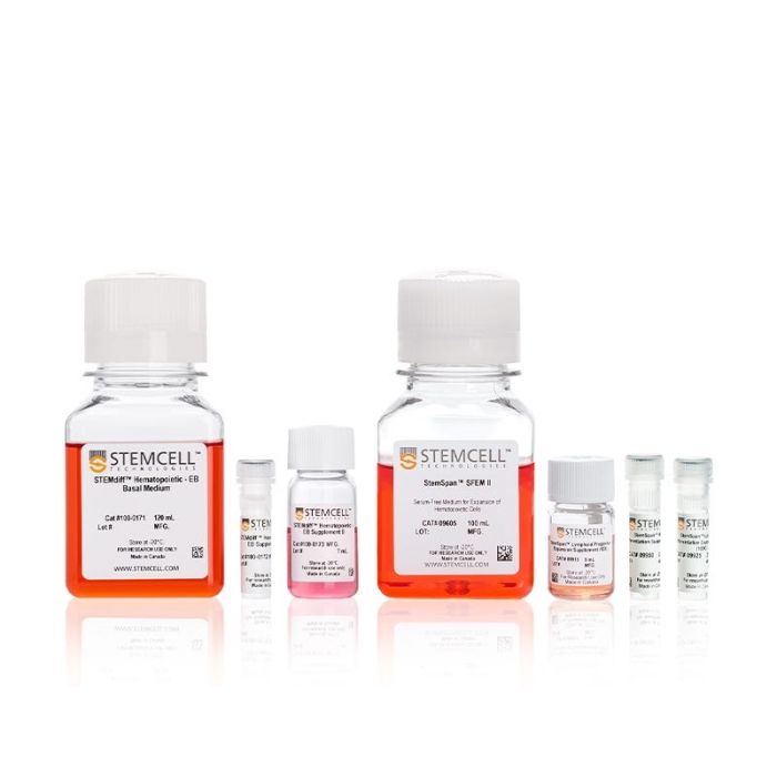
用于hPSC向NK细胞的扩增和分化
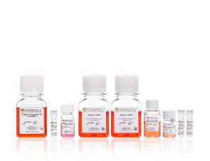
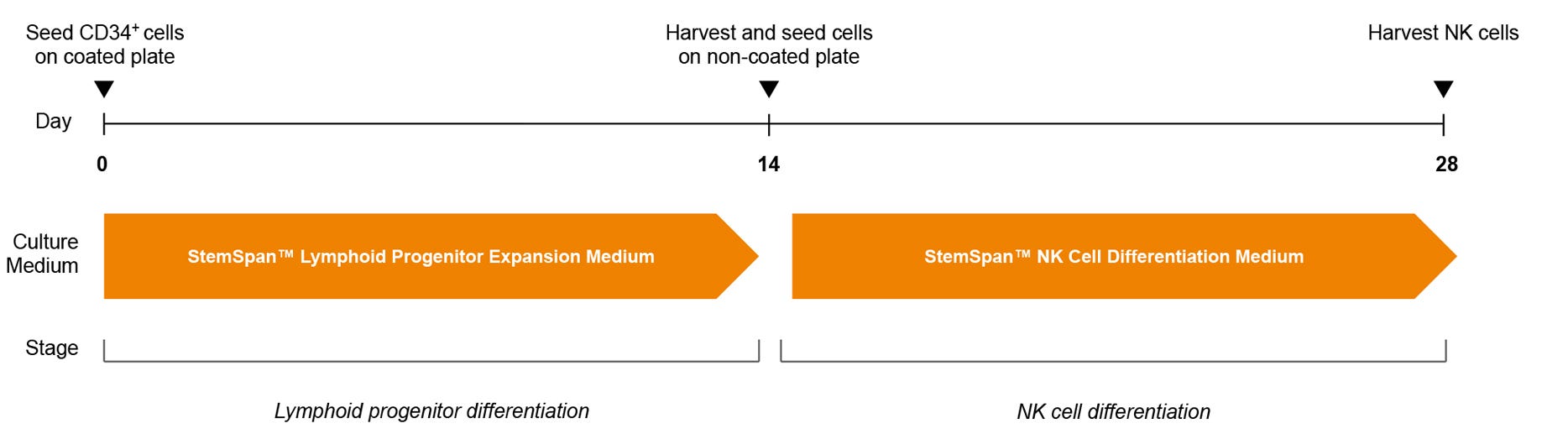
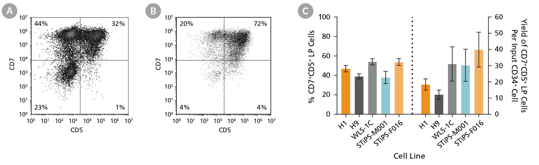
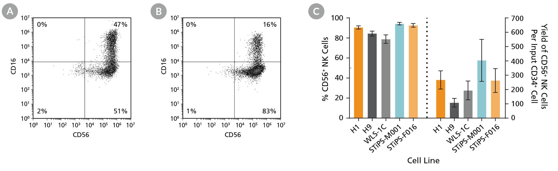
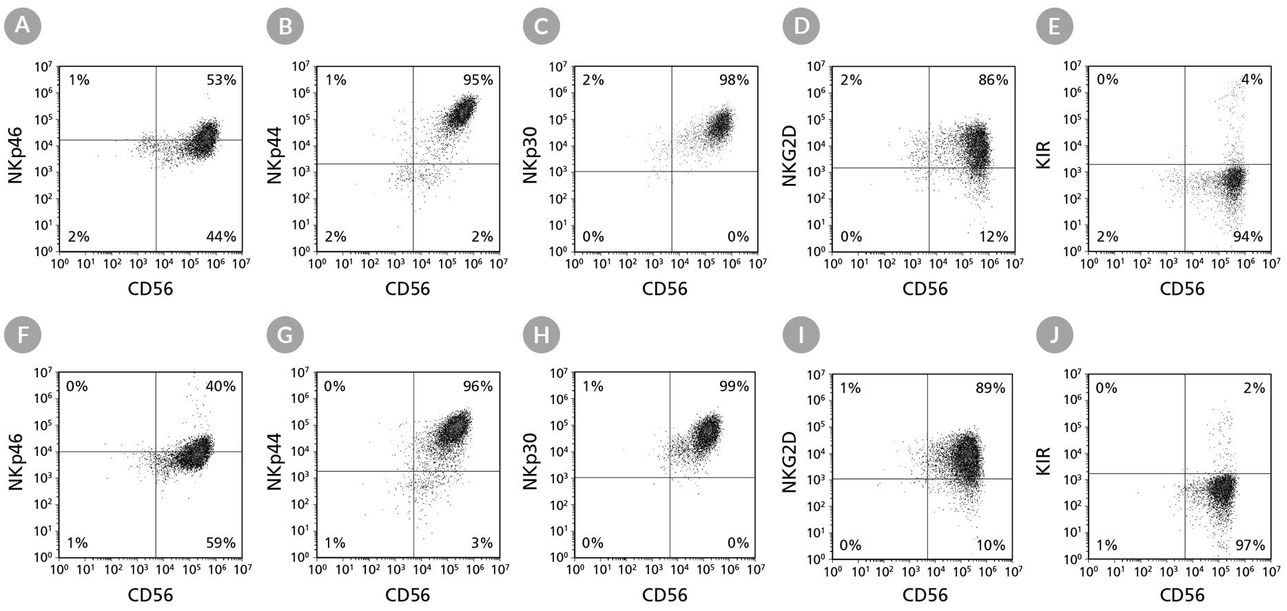
使用无饲养层、无血清的STEMdiff™ NK 细胞分化试剂盒将人多能干细胞(hPSCs)分化为NK细胞
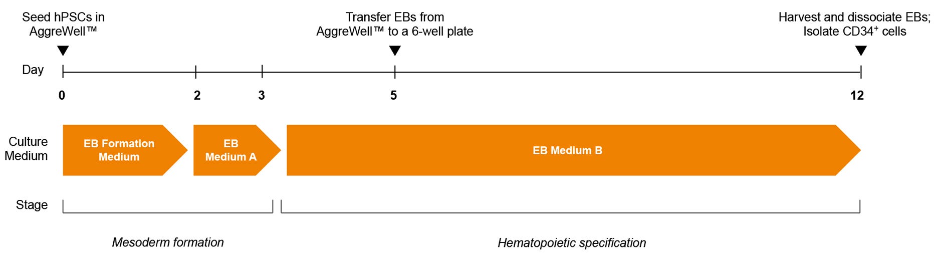
STEMdiff™ NK细胞分化试剂盒方案首先采用无动物成分的STEMdiff™造血分化 - EB试剂,从hPSCs生成拟胚体(EBs),随后进一步分化为CD34+细胞。本试剂盒包含该步骤所需全部EB试剂:
• STEMdiff™造血分化 - EB基础培养基
• STEMdiff™造血分化 - EB补充剂 A
• STEMdiff™造血分化 - EB补充剂 B
随后使用StemSpan™试剂将CD34+细胞进一步定向分化为CD56+ NK细胞:
• StemSpan™ SFEM II
• StemSpan™淋系祖细胞扩增补充剂(10X)
• StemSpan™淋系分化包被材料(100X)
• StemSpan™ NK细胞分化补充剂(100X)
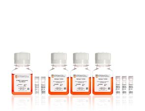
为方便使用,试剂盒内所有STEMdiff™造血分化 - EB及StemSpan™试剂均可单独购买
分类
专用培养基,添加剂
细胞类型
造血细胞,PSC衍生,NK 细胞,多能干细胞
种属
人
应用
细胞培养,分化,扩增
品牌
STEMdiff
研究领域
癌症,疾病建模,药物发现和毒性检测,免疫学,干细胞生物学
制剂类别
无血清
请在《产品说明书》中查找相关支持信息和使用说明,或浏览下方更多实验方案。
本产品专为以下研究领域设计,适用于工作流程中的高亮阶段。探索这些工作流程,了解更多我们为各研究领域提供的其他配套产品。
| 物种 | 人 |
|---|---|
| 配方 | 无血清 |
用于将人多能干细胞(hPSCs)分化为单核细胞
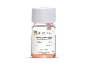
用于人CD34+细胞扩增及分化为淋系祖细胞的添加物
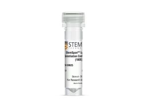
用于淋系祖细胞扩增与分化的包板材料
用于淋系祖细胞向NK细胞分化的添加物
用于生成拟胚体的无血清培养基,以支持下游淋系分化
无动物成分培养补充剂,用于中胚层定向分化并具备下游淋系分化潜能
无动物成分培养补充剂,专为淋系潜能造血分化而优化
用于hPSC向T细胞的扩增和分化
在线联系


 沪公网安备31010102008431号