
 EasySep™小鼠TIL(CD45)正选试剂盒


产品号 #100-0194_C
用于hPSC向T细胞的扩增和分化
若您需要咨询产品或有任何技术问题,请通过官方电话 400 885 9050 或邮箱 info.cn@stemcell.com 与我们联系。
用于hPSC向T细胞的扩增和分化
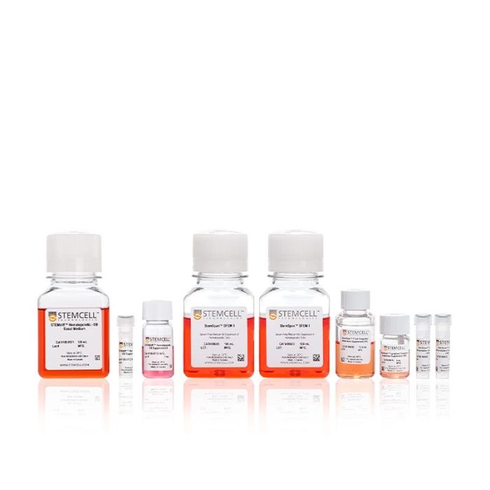
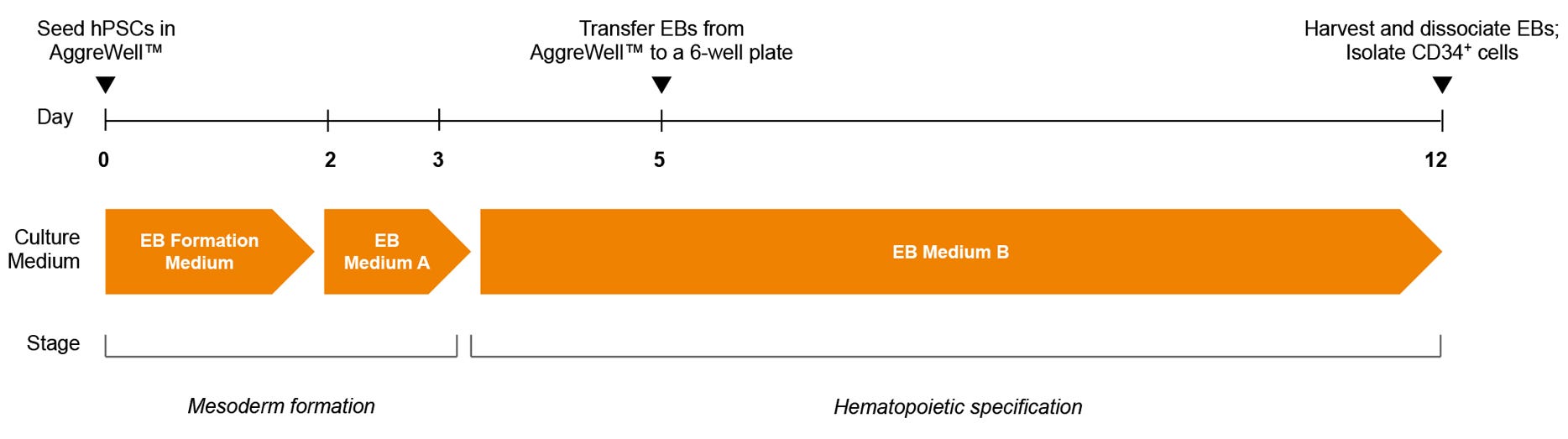
使用无饲养层、无血清的STEMdiff™ T 细胞分化试剂盒将人多能干细胞(hPSCs)分化为T细胞
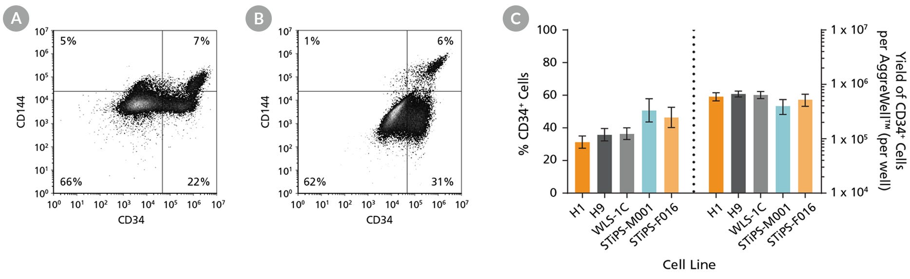
STEMdiff™ T细胞分化试剂盒方案首先采用无动物成分的STEMdiff™造血分化 - EB试剂,从hPSCs生成拟胚体(EBs),随后进一步分化为CD34+细胞。本试剂盒包含该步骤所需全部EB试剂:
• STEMdiff™造血分化 - EB基础培养基
• STEMdiff™造血分化 - EB补充剂 A
• STEMdiff™造血分化 - EB补充剂 B
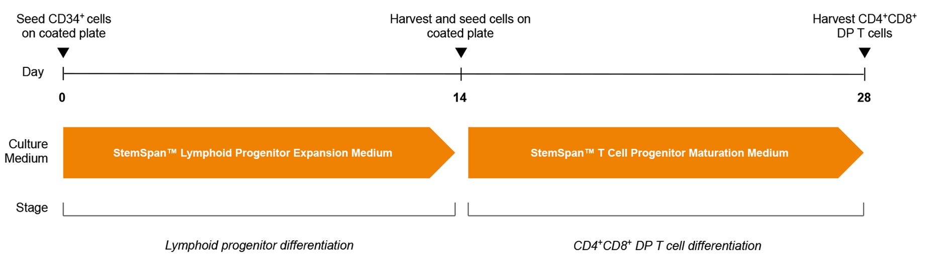
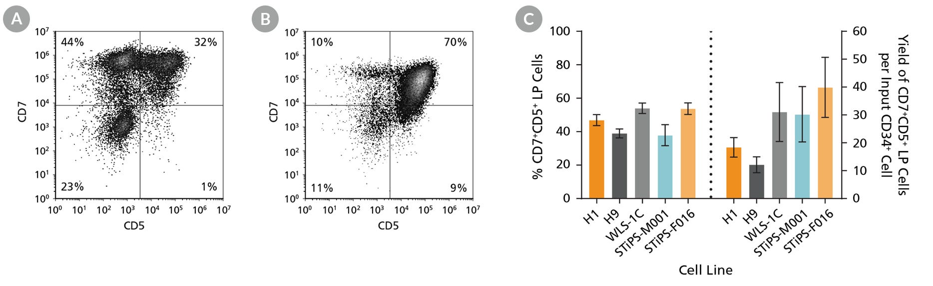
随后使用StemSpan™试剂将CD34+细胞进一步定向分化为T细胞:
• StemSpan™ SFEM II
• StemSpan™淋系祖细胞扩增补充剂(10X)
• StemSpan™淋系分化包被材料(100X)
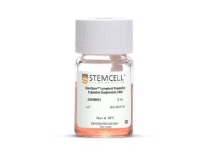
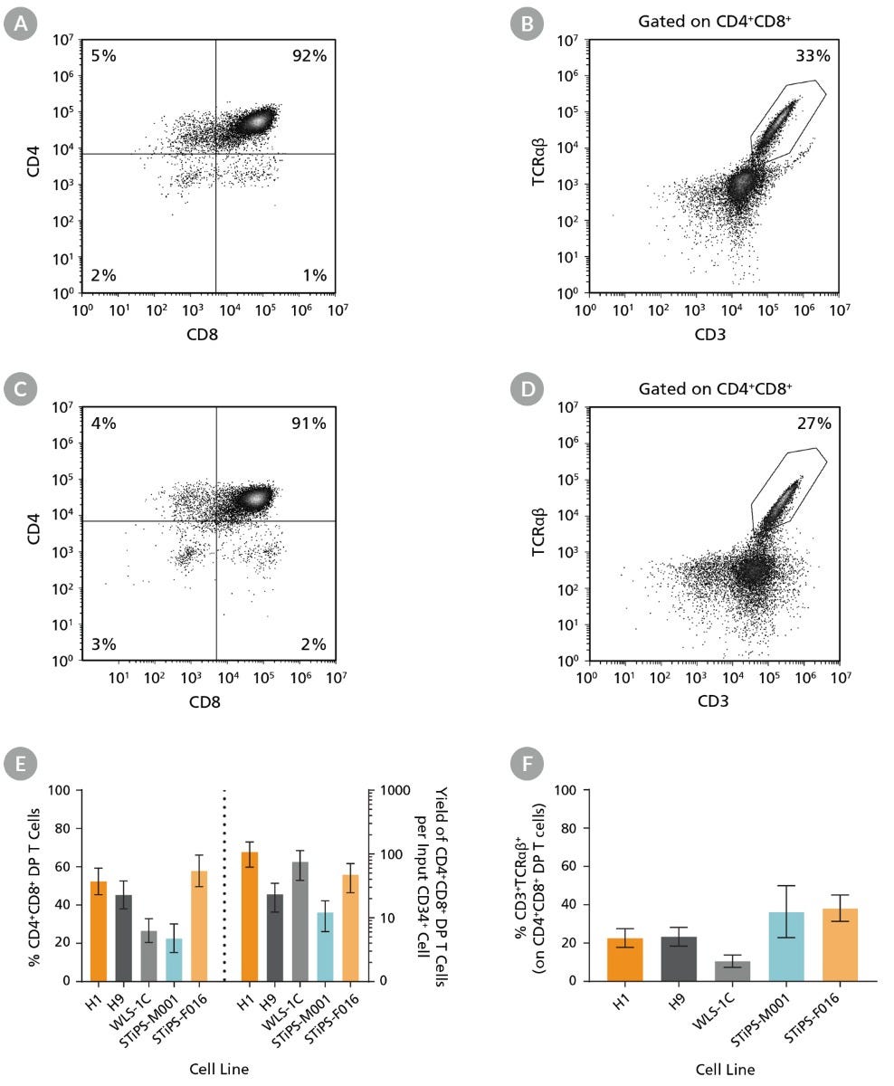
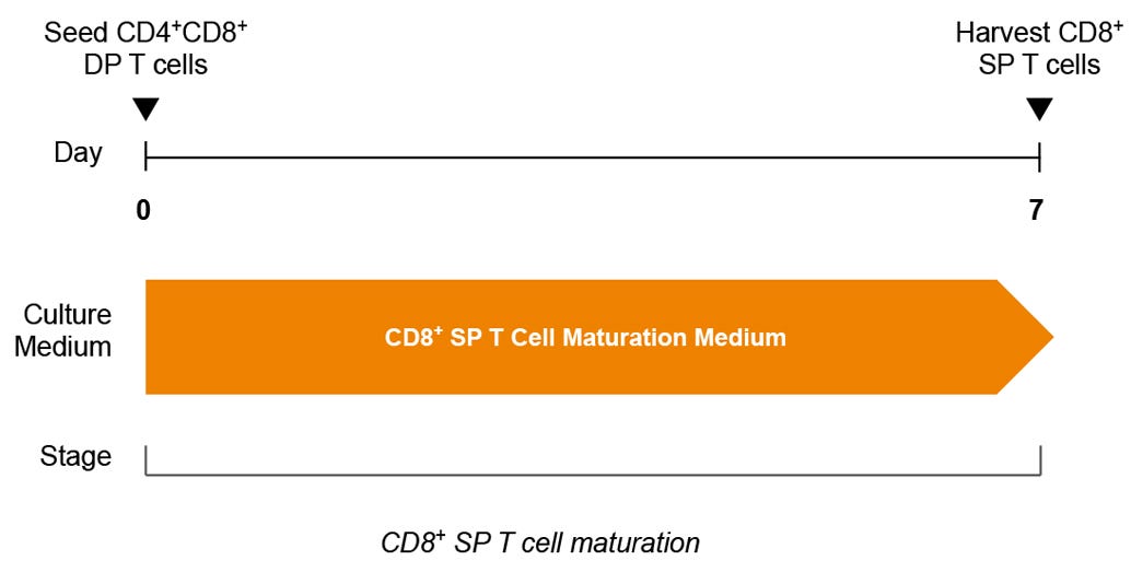
• StemSpan™ T细胞祖细胞成熟添加物(10X)
为方便使用,试剂盒内所有STEMdiff™造血分化 - EB及StemSpan™试剂均可单独购买
分类
专用培养基
细胞类型
造血细胞,PSC衍生,多能干细胞,T 细胞,T 细胞,CD8+
种属
人
应用
细胞培养,分化,扩增
品牌
STEMdiff
研究领域
癌症,疾病建模,药物发现和毒性检测,免疫学,干细胞生物学
制剂类别
无血清
Find supporting information and directions for use in the Product Information Sheet or explore additional protocols below.
This product is designed for use in the following research area(s) as part of the highlighted workflow stage(s). Explore these workflows to learn more about the other products we offer to support each research area.
| 物种 | 人 |
|---|---|
| 配方 | 无血清 |
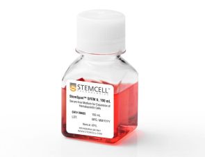
用于培养和扩增造血细胞的无血清培养基
用于人CD34+细胞扩增及分化为淋系祖细胞的添加物
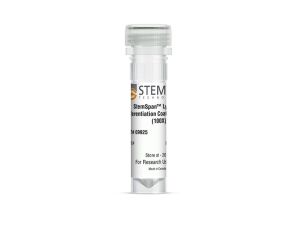
用于淋系祖细胞扩增与分化的包板材料
促进淋巴祖细胞成熟为T细胞的添加物
将人CD34+造血祖细胞扩增并分化为T细胞的全套试剂盒
在线联系


 沪公网安备31010102008431号