
 EasySep™小鼠TIL(CD45)正选试剂盒


搜索结果: 'methocult media formulations for mouse hematopoietic cells serum containing'
-
 抗小鼠IL-4抗体,clone 11B11 抗小鼠IL-4的大鼠Monoclonal 抗体
 -
 抗小鼠Ly-49A抗体,clone YE1/32.8.5 抗小鼠Ly-49A的大鼠Monoclonal IgG2a抗体
 -
 抗小鼠OCT4(OCT3)抗体,clone 40 抗人、鼠OCT4(OCT3)的小鼠Monoclonal IgG1抗体
 -
 抗小鼠TCR Gamma/Delta抗体,clone GL3 亚美尼亚仓鼠Monoclonal IgG2抗体,抗小鼠T细胞受体 gamma/delta
 -
 重组人/小鼠激活素A 激活素A
 -
 重组人/小鼠NT-3 神经营养因子-3
 -
 重组小鼠/大鼠RANTES (CCL5) 激活后调节正常T细胞表达和分泌因子
 -
 EasySep™小鼠/人嵌合体分选试剂盒
对来源于人异种移植受体小鼠的骨髓、脾或外周血样本中未标记人细胞进行免疫磁珠负选分离
-
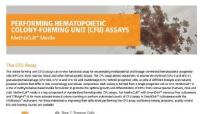 产品手册MethoCult™ Media for Performing Hematopoietic Colony-Forming Unit (CFU) Assays
产品类型:
品牌:
MethoCult
产品号#:
03134
03231
03234
03334
03434
03436
03444
03534
03630
04064
04100
04230
04236
04431
04434
04436
04444
04464
04531
04535
04536
04545
04564
07700
07800
07850
27100
27150
28110
28120
28230
28240
38068
产品名:
MethoCult™M3134
MethoCult™M3231
MethoCult™M3234
MethoCult™M3334
MethoCult™GF M3434
MethoCult™SF M3436
MethoCult™GF M3434
MethoCult™GF M3534
MethoCult™M3630
MethoCult™ H4034 Optimum启动试剂盒套装
MethoCult™ H4100
MethoCult™H4230
MethoCult™SF H4236
MethoCult™H4431
MethoCult™H4434经典
MethoCult™ SF H4436
MethoCult™H4434经典
MethoCult™ H4434 Classic启动试剂盒套装
MethoCult™H4531
MethoCult™H4535富集无EPO
MethoCult™ SF H4536
MethoCult™ H4535 Enriched,不含EPO
入门套件MethoCult™H4534经典无EPO
含 2% FBS的Iscove's MDM
氯化铵溶液
氯化铵溶液
35 mm培养皿
35 mm培养皿
16号钝针
钝针
3 mL注射器
3 mL注射器
Corning® 60 mm 带网格培养皿

 沪公网安备31010102008431号