
 EasySep™小鼠TIL(CD45)正选试剂盒


产品号 #73682_C
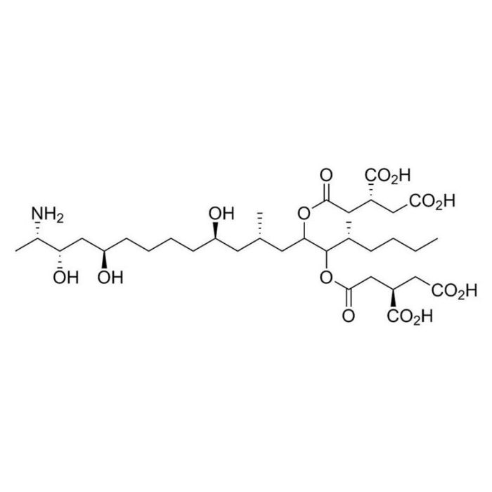
鞘脂合成和蛋白丝氨酸/苏氨酸磷酸酶抑制剂
若您需要咨询产品或有任何技术问题,请通过官方电话 400 885 9050 或邮箱 info.cn@stemcell.com 与我们联系。
Fumonisin B1(伏马菌素B1)是由串珠镰刀菌(Fusarium moniliforme)产生的一种霉菌毒素,已被证明能强效抑制鞘氨醇N-酰基转移酶(神经酰胺合酶;Wang et al.),从而干扰鞘脂(质膜的关键成分)的合成(IC₅₀=0.1 µM)。伏马菌素B1还能抑制蛋白丝氨酸/苏氨酸磷酸酶(PPs;PP1、PP2A、PP2B、PP2C和PP5/T/K/H),IC₅₀值为80-3000 μM。PP5最敏感,IC₅₀值为80 μM(Fukuda et al.)。伏马菌素B1与黄曲霉毒素B1协同作用,可增加大鼠脾细胞的活性氧水平和氧化损伤(Mary et al.)。
维持培养与自我更新
·可逆地阻止瑞士3T3细胞的细胞增殖和DNA合成(Meivar-Levy et al.)。
·阻止十六烷基磷酸胆碱(HePC)诱导的人角质形成细胞系细胞凋亡(Wieder et al.)。
分化
·破坏小脑浦肯野神经元的树突生长(Furuya et al)。
·抑制培养的海马神经元的轴突分支(Schwarz et al.)。
癌症研究
·减弱小鼠淋巴瘤细胞系对血小板活化因子的反应,并通过抑制神经酰胺形成来阻止HePC诱导的细胞凋亡(Balsinde et al.)。
细胞类型
癌细胞及细胞系,角质形成细胞,白血病/淋巴瘤细胞,神经元
种属
人,小鼠,非人灵长类,其他物种,大鼠
应用
分化
研究领域
癌症,神经科学
CAS 编号
116355-83-0
化学式
C₃₄H₅₉NO₁₅
纯度
≥ 95 %
靶点
鞘氨醇N-酰基转移酶
请在《产品说明书》中查找相关支持信息和使用说明,或浏览下方更多实验方案。
| 物种 | 人, 其它物种, 大鼠, 小鼠, 非人灵长类 |
|---|---|
| Cas Number | 116355-83-0 |
| Chemical Formula | C₃₄H₅₉NO₁₅ |
| 纯度 | ≥ 95 % |
| Target | Sphingosine N-Acyltransferase |
培养人神经干细胞和祖细胞的培养基
提升神经元功能的无血清基础培养基
WNT通路激活剂;抑制蛋白磷酸酶PP2A
Hedgehog通路激活剂;激活Smoothened(SMO)
在线联系

 沪公网安备31010102008431号