
 EasySep™小鼠TIL(CD45)正选试剂盒


产品号 #05914_C
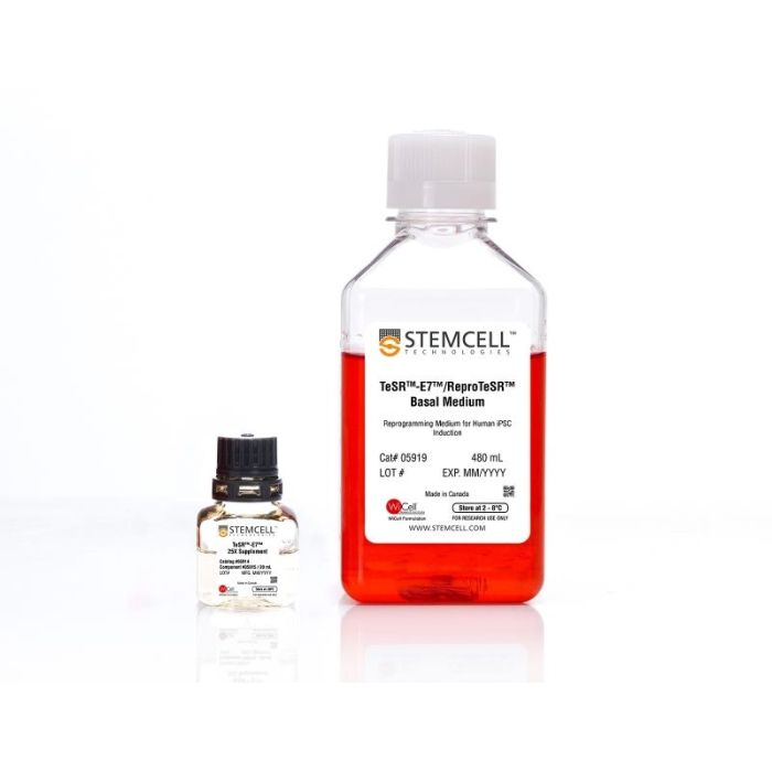
无饲养层且不含动物成分的重编程培养基,用于人诱导性多能干细胞(iPS细胞)的诱导。
若您需要咨询产品或有任何技术问题,请通过官方电话 400 885 9050 或邮箱 info.cn@stemcell.com 与我们联系。
无饲养层且不含动物成分的重编程培养基,用于人诱导性多能干细胞(iPS细胞)的诱导。
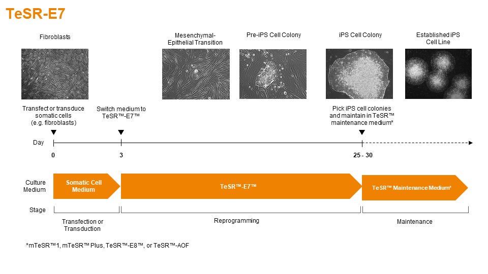
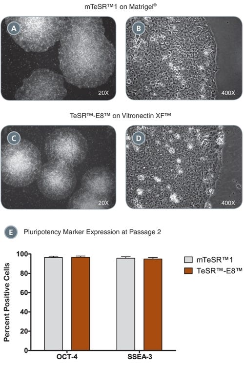
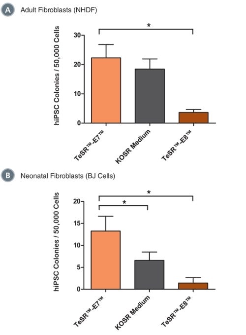
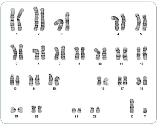
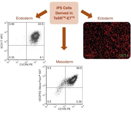
TeSR™-E7™(双组分)是一种不含动物成分、成分明确的重编程培养基,经过优化,旨在生成不依赖饲养层的人诱导性多能干细胞(iPS细胞)。该培养基基于Dr. James Thomson(威斯康星大学麦迪逊分校)实验室发布的E7配方。
分类
专用培养基
细胞类型
多能干细胞
种属
人
应用
细胞培养,重编程
品牌
TeSR
研究领域
干细胞生物学
制剂类别
不含动物成分,无血清,无异源
请在《产品说明书》中查找相关支持信息和使用说明,或浏览下方更多实验方案。
本产品专为以下研究领域设计,适用于工作流程中的高亮阶段。探索这些工作流程,了解更多我们为各研究领域提供的其他配套产品。
| 物种 | 人类 |
|---|---|
| 配方 | 不含动物成分, 无血清 |
无饲养层、无动物成分的人胚胎干细胞和诱导多能干细胞维持培养基
成分明确的无异源基质,支持人多能干细胞在无血清、无饲养层条件下的生长和分化。
<p>cGMP标准、无饲养层的hESC和iPSC维持培养基</p>
在线联系


 沪公网安备31010102008431号