产品号 #05914_C
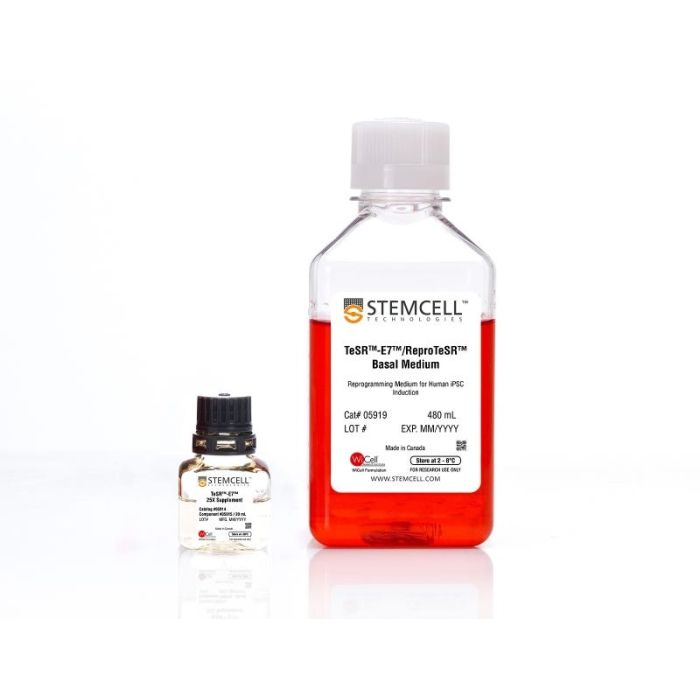
无饲养层且不含动物成分的重编程培养基,用于人诱导性多能干细胞(iPS细胞)的诱导。
若您需要咨询产品或有任何技术问题,请通过官方电话 400 885 9050 或邮箱 info.cn@stemcell.com 与我们联系。
无饲养层且不含动物成分的重编程培养基,用于人诱导性多能干细胞(iPS细胞)的诱导。
无饲养层且不含动物成分的重编程培养基,用于人诱导性多能干细胞(iPS细胞)的诱导。
TeSR™-E7™(双组分)是一种不含动物成分、成分明确的重编程培养基,经过优化,旨在生成不依赖饲养层的人诱导性多能干细胞(iPS细胞)。该培养基基于Dr. James Thomson(威斯康星大学麦迪逊分校)实验室发布的E7配方。
分类
专用培养基
细胞类型
多能干细胞
种属
人
应用
细胞培养,重编程
品牌
TeSR
研究领域
干细胞生物学
制剂类别
不含动物成分,无血清,无异源
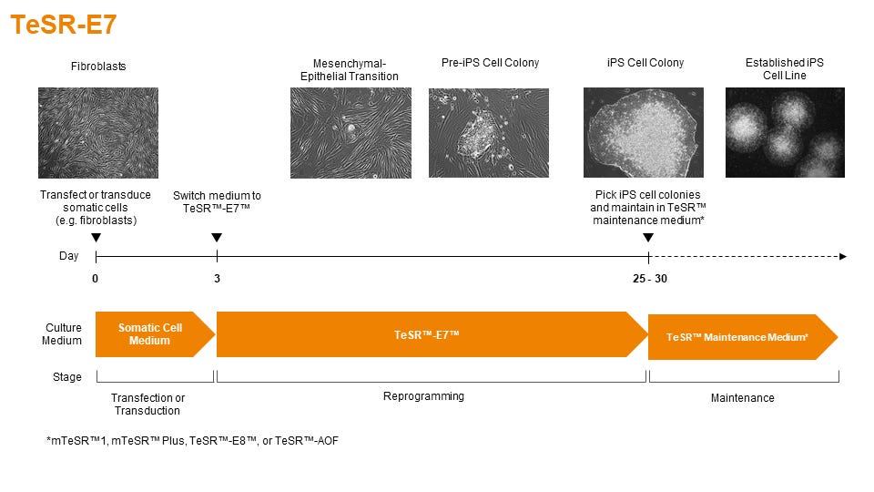
Figure 1. Schematic of Reprogramming Timeline
TeSR™-E7™ can be used during the entire induction phase of reprogramming (day 3 to 25+). Following reprogramming, iPS cell colonies can be isolated and propogated in feeder-free maintenance systems (eg. mTeSR™1 or TeSR™-E8™ media on Corning® Matrigel® or Vitronectin XF™ matrices).
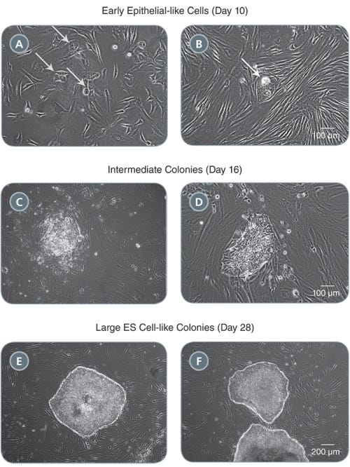
Figure 2. Morphology of Representative iPS Cell Colonies Arising During the Induction Period in TeSR™-E7™
(A-B) Small clusters of colonies with an epithelial-like morphology will appear by one to two weeks following induction (see arrows). (C-D) These clusters expand into pre-iPS cell colonies by two to three weeks. (E-F) Larger ES cell-like colonies are clearly identifiable by three to four weeks. Representative colonies from adult human fibroblasts reprogrammed with episomal vectors containing OCT-4, SOX2, KLF-4, and L-MYC are shown.
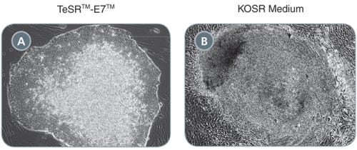
Figure 3. Comparison of Primary iPS Cell Colonies Derived Using TeSR™-E7™ and KOSR-Based Medium
(A) TeSR™-E7™ generates colonies with defined borders and less overgrowth of background fibroblasts compared to (B) KOSR-based iPS cell induction medium. Representative colonies from adult human fibroblasts reprogrammed with episomal vectors containing OCT-4, SOX2, KLF-4, and L-MYC are shown.
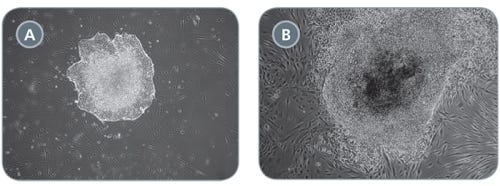
Figure 4. Comparison of Primary iPS Cell Colonies Derived Using TeSR™-E7™ with Qualified vs Unqualified bFGF
(A) TeSR™-E7™ yields easily recognizable iPS cell colonies with defined borders. (B) Unqualified components can result in colonies that have poorly defined edges and higher levels of differentiation. Representative colonies from adult human fibroblasts reprogrammed with episomal vectors containing OCT-4, SOX2, KLF-4, and L-MYC are shown.
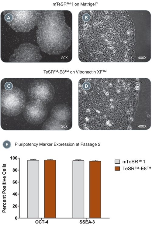
Figure 5. iPS Colonies Expanded in mTeSR™ or TeSR™-E8™
(A - D) iPS cell colonies generated in TeSR™-E7™ and expanded in either mTeSR™1 on Corning® Matrigel® (A-B) or TeSR™-E8™ on Vitronectin XF™ (C, D) exhibit classic ES cell morphology with dense colony centers, defined borders, prominent nucleoli and high nuclear-to-cytoplasmic ratios. (E) iPS cells express high levels of pluripotency markers after just two passages in either mTeSR™1 or TeSR™-E8™ as demonstrated by OCT-4 and SSEA-3 flow cytometry analysis. Data are expressed as mean ± SEM, n = 4.
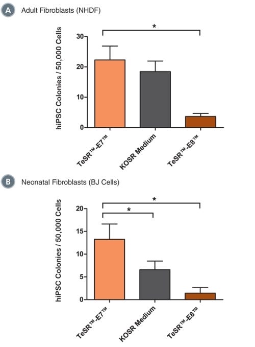
Figure 6. TeSR™-E7™ Supports Reprogramming of Human Cell Types Including Adult Dermal Fibroblasts and Neonatal Fibroblasts
Reprogramming of (A) adult normal human dermal fibroblasts (NHDF, 33 year-old female) and (B) neonatal foreskin fibroblasts (BJ cells) with episomal reprogramming vectors are shown. TeSR™-E7™ demonstrated similar (in NHDF) or greater (in BJ cells) reprogramming efficiencies compared to KOSR-based iPS cell induction medium. TeSR™-E7™ demonstrated higher reprogramming efficiencies compared to TeSR™-E8™. Data are expressed as mean ± SEM, n ≥ 6, * p ≤ 0.05.
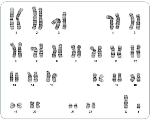
Figure 7. iPS Cells Derived in TeSR™-E7™ Display Normal Karyotype
iPS cell lines were generated in TeSR™-E7™ medium, maintained in mTeSR™1 or TeSR™-E8™ media for a minimum of 5 passages and karyotyped by G-banding karyotype analysis. Three iPS cell lines were analyzed and all demonstrated a normal karyotype; a representative karyogram is shown.
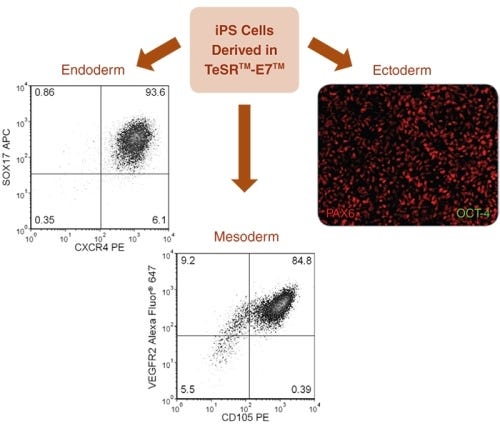
Figure 8. Directed Differentiation of iPS Cells to All Three Germ Layers
TeSR™-E7™-derived iPS cells were differentiated into all three germ layers. Endoderm specification was achieved using the STEMdiff™ Definitive Endoderm Kit, results demonstrated 93.6% SOX17 + CXCR4 + cells. Mesoderm specification was demonstrated using a STEMdiff™ APEL™ medium-based endothelial differentiation protocol, results demonstrated &ht;99% CD31 + cells (data not shown) and 84.8% VEGFR2 + CD105 + cells. Ectoderm specification was demonstrated using STEMdiff™ Neural Induction Medium, immunocytochemistry shows high levels of PAX6 staining with no detectable OCT-4 staining by day 9 of neural induction.
请在《产品说明书》中查找相关支持信息和使用说明,或浏览下方更多实验方案。
本产品专为以下研究领域设计,适用于工作流程中的高亮阶段。探索这些工作流程,了解更多我们为各研究领域提供的其他配套产品。
Thank you for your interest in IntestiCult™ Organoid Growth Medium (Human). Please provide us with your contact information and your local representative will contact you with a customized quote. Where appropriate, they can also assist you with a(n):
Estimated delivery time for your area
Product sample or exclusive offer
In-lab demonstration
| 物种 | 人类 |
|---|---|
| 配方 | 不含动物成分, 无血清 |
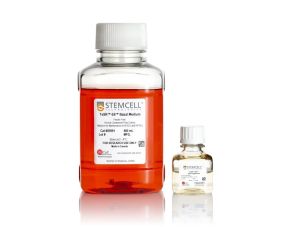
无饲养层、无动物成分的人胚胎干细胞和诱导多能干细胞维持培养基
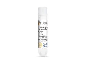
成分明确的无异源基质,支持人多能干细胞在无血清、无饲养层条件下的生长和分化。
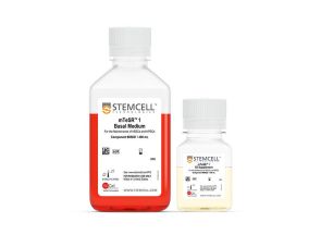
<p>cGMP标准、无饲养层的hESC和iPSC维持培养基</p>
扫描二维码或搜索微信号STEMCELLTech,即可关注我们的微信平台,第一时间接收丰富的技术资源和最新的活动信息。
如您有任何问题,欢迎发消息给STEMCELLTech微信公众平台,或与我们通过电话/邮件联系:400 885 9050 INFO.CN@STEMCELL.COM。
在线联系

