Kourjian G et al. (MAY 2016)
Journal of Immunology 196 9 3595--607
HIV Protease Inhibitor-Induced Cathepsin Modulation Alters Antigen Processing and Cross-Presentation.
Immune recognition by T cells relies on the presentation of pathogen-derived peptides by infected cells,but the persistence of chronic infections calls for new approaches to modulate immune recognition. Ag cross-presentation,the process by which pathogen Ags are internalized,degraded,and presented by MHC class I,is crucial to prime CD8 T cell responses. The original degradation of Ags is performed by pH-dependent endolysosomal cathepsins. In this article,we show that HIV protease inhibitors (PIs) prescribed to HIV-infected persons variably modulate cathepsin activities in human APCs,dendritic cells and macrophages,and CD4 T cells,three cell subsets infected by HIV. Two HIV PIs acted in two complementary ways on cathepsin hydrolytic activities: directly on cathepsins and indirectly on their regulators by inhibiting Akt kinase activities,reducing NADPH oxidase 2 activation,and lowering phagolysosomal reactive oxygen species production and pH,which led to enhanced cathepsin activities. HIV PIs modified endolysosomal degradation and epitope production of proteins from HIV and other pathogens in a sequence-dependent manner. They altered cross-presentation of Ags by dendritic cells to epitope-specific T cells and T cell-mediated killing. HIV PI-induced modulation of Ag processing partly changed the MHC self-peptidome displayed by primary human cells. This first identification,to our knowledge,of prescription drugs modifying the regulation of cathepsin activities and the MHC-peptidome may provide an alternate therapeutic approach to modulate immune recognition in immune disease beyond HIV.
View Publication
产品号#:
17952
17952RF
100-0696
19654
19654RF
产品名:
EasySep™人CD4+ T细胞分选试剂盒
RoboSep™ 人CD4+ T细胞分选试剂盒
EasySep™人CD4+ T细胞分离试剂盒
EasySep™ Direct 人 PBMC 分选试剂盒
RoboSep™ Direct 人 PBMC 分选试剂盒
Hrecka K et al. (JUL 2016)
Proceedings of the National Academy of Sciences of the United States of America 113 27 E3921--30
HIV-1 and HIV-2 exhibit divergent interactions with HLTF and UNG2 DNA repair proteins.
HIV replication in nondividing host cells occurs in the presence of high concentrations of noncanonical dUTP,apolipoprotein B mRNA-editing,enzyme-catalytic,polypeptide-like 3 (APOBEC3) cytidine deaminases,and SAMHD1 (a cell cycle-regulated dNTP triphosphohydrolase) dNTPase,which maintains low concentrations of canonical dNTPs in these cells. These conditions favor the introduction of marks of DNA damage into viral cDNA,and thereby prime it for processing by DNA repair enzymes. Accessory protein Vpr,found in all primate lentiviruses,and its HIV-2/simian immunodeficiency virus (SIV) SIVsm paralogue Vpx,hijack the CRL4(DCAF1) E3 ubiquitin ligase to alleviate some of these conditions,but the extent of their interactions with DNA repair proteins has not been thoroughly characterized. Here,we identify HLTF,a postreplication DNA repair helicase,as a common target of HIV-1/SIVcpz Vpr proteins. We show that HIV-1 Vpr reprograms CRL4(DCAF1) E3 to direct HLTF for proteasome-dependent degradation independent from previously reported Vpr interactions with base excision repair enzyme uracil DNA glycosylase (UNG2) and crossover junction endonuclease MUS81,which Vpr also directs for degradation via CRL4(DCAF1) E3. Thus,separate functions of HIV-1 Vpr usurp CRL4(DCAF1) E3 to remove key enzymes in three DNA repair pathways. In contrast,we find that HIV-2 Vpr is unable to efficiently program HLTF or UNG2 for degradation. Our findings reveal complex interactions between HIV-1 and the DNA repair machinery,suggesting that DNA repair plays important roles in the HIV-1 life cycle. The divergent interactions of HIV-1 and HIV-2 with DNA repair enzymes and SAMHD1 imply that these viruses use different strategies to guard their genomes and facilitate their replication in the host.
View Publication
产品号#:
19052
19052RF
19051
19051RF
产品名:
EasySep™人CD4+ T细胞富集试剂盒
RoboSep™ 人CD4+ T细胞富集试剂盒含滤芯吸头
EasySep™人T细胞富集试剂盒
RoboSep™ 人T细胞富集试剂盒含滤芯吸头
Carroll VA et al. (OCT 2016)
Proceedings of the National Academy of Sciences of the United States of America
Expression of HIV-1 matrix protein p17 and association with B-cell lymphoma in HIV-1 transgenic mice.
HIV-1 infection is associated with increased risk for B-cell lymphomas. How HIV infection promotes the development of lymphoma is unclear,but it may involve chronic B-cell activation,inflammation,and/or impaired immunity,possibly leading to a loss of control of oncogenic viruses and reduced tumor immunosurveillance. We hypothesized that HIV structural proteins may contribute to lymphomagenesis directly,because they can persist long term in lymph nodes in the absence of viral replication. The HIV-1 transgenic mouse Tg26 carries a noninfectious HIV-1 provirus lacking part of the gag-pol region,thus constituting a model for studying the effects of viral products in pathogenesis. Approximately 15% of Tg26 mice spontaneously develop leukemia/lymphoma. We investigated which viral proteins are associated with the development of leukemia/lymphoma in the Tg26 mouse model,and performed microarray analysis on RNA from spleen and lymph nodes to identify potential mechanisms of lymphomagenesis. Of the viral proteins examined,only expression of HIV-1 matrix protein p17 was associated with leukemia/lymphoma development and was highly expressed in bone marrow before disease. The tumor cells resembled pro-B cells,and were CD19(+)IgM(-)IgD(-)CD93(+)CD43(+)CD21(-)CD23(-)VpreB(+)CXCR4(+) Consistent with the pro-B-cell stage of B-cell development,microarray analysis revealed enrichment of transcripts,including Rag1,Rag2,CD93,Vpreb1,Vpreb3,and Igll1 We confirmed RAG1 expression in Tg26 tumors,and hypothesized that HIV-1 matrix protein p17 may directly induce RAG1 in B cells. Stimulation of human activated B cells with p17 enhanced RAG1 expression in three of seven donors,suggesting that intracellular signaling by p17 may lead to genomic instability and transformation.
View Publication
产品号#:
19054
19054RF
17963
17963RF
产品名:
EasySep™人B细胞富集试剂盒
RoboSep™ 人B细胞富集试剂盒含滤芯吸头
EasySep™人B细胞富集试剂盒II(不去除CD43)
RoboSep™ 人B细胞富集试剂盒II(不去除CD43)
Vanwalscappel B et al. (NOV 2016)
Virology 500 247--258
Genetic and phenotypic analyses of sequential vpu alleles from HIV-infected IFN-treated patients.
Treatment of HIV-infected patients with IFN-α results in significant,but clinically insufficient,reductions of viremia. IFN induces the expression of several antiviral proteins including BST-2,which inhibits HIV by multiple mechanisms. The viral protein Vpu counteracts different effects of BST-2. We thus asked if Vpu proteins from IFN-treated patients displayed improved anti-BST-2 activities as compared to Vpu from baseline. Deep-sequencing analyses revealed that in five of seven patients treated by IFN-α for a concomitant HCV infection in the absence of antiretroviral drugs,the dominant Vpu sequences differed before and during treatment. In three patients,vpu alleles that emerged during treatment improved virus replication in the presence of IFN-α,and two of them conferred improved virus budding from cells expressing BST-2. Differences were observed for the ability to down-regulate CD4,while all Vpu variants potently down-modulated BST-2 from the cell surface. This report discloses relevant consequences of IFN-treatment on HIV properties.
View Publication
产品号#:
19052
19052RF
85450
85460
86450
86460
产品名:
EasySep™人CD4+ T细胞富集试剂盒
RoboSep™ 人CD4+ T细胞富集试剂盒含滤芯吸头
SepMate™-50 (IVD)
SepMate™-50 (IVD)
SepMate™-50 (RUO)
SepMate™-50 (RUO)
Cesaro A et al. (SEP 2012)
PLoS ONE 7 9 e45478
An inflammation loop orchestrated by S100A9 and Calprotectin is critical for development of arthritis
OBJECTIVE: The S100A9 and S100A8 proteins are highly expressed by neutrophils and monocytes and are part of a group of damage-associated molecular pattern molecules that trigger inflammatory responses. Sera and synovial fluids of patients with rheumatoid arthritis (RA) contain high concentrations of S100A8/A9 that correlate with disease activity.backslashnbackslashnMETHODS: In this study,we investigated the importance of S100A9 in RA by using neutralizing antibodies in a murine lipopolysaccharide-synchronized collagen-induced arthritis model. We also used an in vitro model of stimulation of human immune cells to decipher the role played by S100A9 in leukocyte migration and pro-inflammatory cytokine secretion.backslashnbackslashnRESULTS: Treatment with anti-S100A9 antibodies improved the clinical score by 50%,diminished immune cell infiltration,reduced inflammatory cytokines,both in serum and in the joints,and preserved bone/collagen integrity. Stimulation of neutrophils with S100A9 protein led to the enhancement of neutrophil transendothelial migration. S100A9 protein also induced the secretion by monocytes of proinflammatory cytokines like TNFα,IL-1β and IL-6,and of chemokines like MIP-1α and MCP-1.backslashnbackslashnCONCLUSION: The effects of anti-S100A9 treatment are likely direct consequences of inhibiting the S100A9-mediated promotion of neutrophil transmigration and secretion of pro-inflammatory cytokines from monocytes. Collectively,our results show that treatment with anti-S100A9 may inhibit amplification of the immune response and help preserve tissue integrity. Therefore,S100A9 is a promising potential therapeutic target for inflammatory diseases like rheumatoid arthritis for which alternative therapeutic strategies are needed.
View Publication
产品号#:
03800
03801
03802
03803
03804
03805
03806
15028
15068
产品名:
ClonaCell™-HY杂交瘤试剂盒
ClonaCell™-HY培养基A
ClonaCell™-HY 培养基 B
ClonaCell™-HY 培养基 C
ClonaCell™-HY 培养基 D
ClonaCell™-HY 培养基 E
ClonaCell™-HY PEG
RosetteSep™人单核细胞富集抗体混合物
RosetteSep™人单核细胞富集抗体混合物
Trkola A et al. (DEC 2003)
Journal of virology 77 24 13146--55
Human immunodeficiency virus type 1 fitness is a determining factor in viral rebound and set point in chronic infection.
Human immunodeficiency virus type 1 (HIV-1) isolates from 20 chronically infected patients who participated in a structured treatment interruption (STI) trial were studied to determine whether viral fitness influences reestablishment of viremia. Viruses derived from individuals who spontaneously controlled viremia had significantly lower in vitro replication capacities than viruses derived from individuals that did not control viremia after interruption of antiretroviral therapy (ART),and replication capacities correlated with pre-ART and post-STI viral set points. Of note,no clinically relevant improvement of viral loads upon STI occurred. Virus isolates from controlling and noncontrolling patients were indistinguishable in terms of coreceptor usage,genetic subtype,and sensitivity to neutralizing antibodies. In contrast,viruses from controlling patients exhibited increased sensitivity to inhibition by chemokines. Sensitivity to inhibition by RANTES correlated strongly with slower replication kinetics of the virus isolates,suggesting a marked dependency of these virus isolates on high coreceptor densities on the target cells. In summary,our data indicate that viral fitness is a driving factor in determining the magnitude of viral rebound and viral set point in chronic HIV-1 infection,and thus fitness should be considered as a parameter influencing the outcome of therapeutic intervention in chronic infection.
View Publication
Addo MM et al. (FEB 2003)
Journal of virology 77 3 2081--92
Comprehensive epitope analysis of human immunodeficiency virus type 1 (HIV-1)-specific T-cell responses directed against the entire expressed HIV-1 genome demonstrate broadly directed responses, but no correlation to viral load.
Cellular immune responses play a critical role in the control of human immunodeficiency virus type 1 (HIV-1); however,the breadth of these responses at the single-epitope level has not been comprehensively assessed. We therefore screened peripheral blood mononuclear cells (PBMC) from 57 individuals at different stages of HIV-1 infection for virus-specific T-cell responses using a matrix of 504 overlapping peptides spanning all expressed HIV-1 proteins in a gamma interferon-enzyme-linked immunospot (Elispot) assay. HIV-1-specific T-cell responses were detectable in all study subjects,with a median of 14 individual epitopic regions targeted per person (range,2 to 42),and all 14 HIV-1 protein subunits were recognized. HIV-1 p24-Gag and Nef contained the highest epitope density and were also the most frequently recognized HIV-1 proteins. The total magnitude of the HIV-1-specific response ranged from 280 to 25,860 spot-forming cells (SFC)/10(6) PBMC (median,4,245) among all study participants. However,the number of epitopic regions targeted,the protein subunits recognized,and the total magnitude of HIV-1-specific responses varied significantly among the tested individuals,with the strongest and broadest responses detectable in individuals with untreated chronic HIV-1 infection. Neither the breadth nor the magnitude of the total HIV-1-specific CD8+-T-cell responses correlated with plasma viral load. We conclude that a peptide matrix-based Elispot assay allows for rapid,sensitive,specific,and efficient assessment of cellular immune responses directed against the entire expressed HIV-1 genome. These data also suggest that the impact of T-cell responses on control of viral replication cannot be explained by the mere quantification of the magnitude and breadth of the CD8+-T-cell response,even if a comprehensive pan-genome screening approach is applied.
View Publication
产品号#:
15022
15062
15023
15063
产品名:
RosetteSep™人CD4+ T细胞富集抗体混合物
RosetteSep™人CD4+ T细胞富集抗体混合物
RosetteSep™人CD8+ T细胞富集抗体混合物
RosetteSep™人CD8+ T细胞富集抗体混合物
Lin S et al. (SEP 2010)
Journal of virology 84 18 9487--96
HIV infection upregulates caveolin 1 expression to restrict virus production.
Caveolin 1 (Cav-1) is a major protein of a specific membrane lipid raft known as caveolae. Cav-1 interacts with the gp41 of the human immunodeficiency virus (HIV) envelope,but the role of Cav-1 in HIV replication and pathogenesis is not known. In this report,we demonstrate that HIV infection in primary human monocyte-derived macrophages (MDMs),THP-1 macrophages,and U87-CD4 cells results in a dramatic upregulation of Cav-1 expression mediated by HIV Tat. The activity of p53 is essential for Tat-induced Cav-1 expression,as our findings show enhanced phosphorylation of serine residues at amino acid positions 15 and 46 in the presence of Tat with a resulting Cav-1 upregulation. Furthermore,inhibition of p38 mitogen-activated protein kinase (MAPK) blocked phosphorylation of p53 in the presence of Tat. Infection studies of Cav-1-overexpressing cells reveal a significant reduction of HIV production. Taken together,these results suggest that HIV infection enhances the expression of Cav-1,which subsequently causes virus reduction,suggesting that Cav-1 may contribute to persistent infection in macrophages.
View Publication
产品号#:
19058
19058RF
100-1525
19059
19059RF
产品名:
EasySep™人单核细胞富集试剂盒(不去除CD16)
RoboSep™ 人单核细胞富集试剂盒(不去除CD16)含滤芯吸头
EasySep™人单核细胞富集试剂盒(不去除CD16)
EasySep™人单核细胞富集试剂盒
RoboSep™ 人单核细胞富集试剂盒含滤芯吸头
Wu X et al. (DEC 2008)
Blood 112 12 4675--82
Alternative splicing regulates activation-induced cytidine deaminase (AID): implications for suppression of AID mutagenic activity in normal and malignant B cells.
The mutagenic enzyme activation-induced cytidine deaminase (AID) is required for immunoglobulin class switch recombination (CSR) and somatic hypermutation (SHM) in germinal center (GC) B cells. Deregulated expression of AID is associated with various B-cell malignancies and,currently,it remains unclear how AID activity is extinguished to avoid illegitimate mutations. AID has also been shown to be alternatively spliced in malignant B cells,and there is limited evidence that this also occurs in normal blood B cells. The functional significance of these splice variants remains unknown. Here we show that normal GC human B cells and blood memory B cells similarly express AID splice variants and show for the first time that AID splicing variants are singly expressed in individual normal B cells as well as malignant B cells from chronic lymphocytic leukemia patients. We further demonstrate that the alternative AID splice variants display different activities ranging from inactivation of CSR to inactivation or heightened SHM activity. Our data therefore suggest that CSR and SHM are differentially switched off by varying the expression of splicing products of AID at the individual cell level. Most importantly,our findings suggest a novel tumor suppression mechanism by which unnecessary AID mutagenic activities are promptly contained for GC B cells.
View Publication
产品号#:
21000
20119
20155
19054
19054RF
19754
19754RF
产品名:
RoboSep™- S
RoboSep™ 吸头组件抛光剂
RoboSep™分选管套装(9个塑料管)
EasySep™人B细胞富集试剂盒
RoboSep™ 人B细胞富集试剂盒含滤芯吸头
Kootstra NA et al. (FEB 2003)
Proceedings of the National Academy of Sciences of the United States of America 100 3 1298--303
Abrogation of postentry restriction of HIV-1-based lentiviral vector transduction in simian cells.
HIV-1 replication in simian cells is restricted at an early postentry step because of the presence of an inhibitory cellular factor. This block reduces the usefulness of HIV-1-based lentiviral vectors in primate animal models. Here,we demonstrate that substitution of the cyclophilin A (CyPA) binding region in the capsid of an HIV-1-based lentiviral vector (LV) with that of the macrophage tropic HIV-1 Ba-L resulted in a vector that was resistant to the inhibitory effect and efficiently transduced simian cells. Notably,the chimeric gag LV efficiently transduced primary simian hematopoietic progenitor cells,a critical cellular target in gene therapy. The alterations in the CyPA binding region did not affect CyPA incorporation; however,transduction by the gag chimeric LV seemed to be relatively insensitive to cyclosporin A,indicating that it does not require CyPA for early postentry steps. In dual infection experiments,the gag chimeric LV failed to remove the block to transduction of the WT LV,suggesting that the gag chimeric LV did not saturate the inhibitory simian cellular factor. These data suggest that the CyPA binding region of capsid contains a viral determinant involved in the postentry restriction of HIV-1-based lentiviral vectors. Overall,the findings demonstrate that the host range of HIV-1-based LV can be altered by modifications in the packaging construct.
View Publication

 EasySep™小鼠TIL(CD45)正选试剂盒


 挂图The Immune Response to HIV Poster Summary of how HIV subverts the immune response to establish a chronic infection
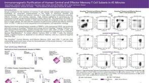 科学海报Immunomagnetic Purification of Human Central and Effector Memory T Cell Subsets in 45 Minutes
 沪公网安备31010102008431号