
 EasySep™小鼠TIL(CD45)正选试剂盒


技术资料
-
 31:49
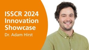
线上讲座Using Human Pluripotent Stem Cell-Derived Neural Organoids for Disease Modeling发布日期: 08/01/2024
31:49
线上讲座Using Human Pluripotent Stem Cell-Derived Neural Organoids for Disease Modeling发布日期: 08/01/2024 -
 24:25
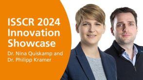
线上讲座ISSCR Standards in Action: Advancements in Cell Line Generation and Organoid Innovation发布日期: 08/01/2024
24:25
线上讲座ISSCR Standards in Action: Advancements in Cell Line Generation and Organoid Innovation发布日期: 08/01/2024 -
 57:26
线上讲座Consequences of Culture-Acquired Genetic Changes in Human Pluripotent Stem Cells发布日期: 02/20/2024
57:26
线上讲座Consequences of Culture-Acquired Genetic Changes in Human Pluripotent Stem Cells发布日期: 02/20/2024 -
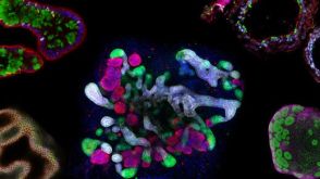 研究综述The Predictive Power of Organoid-Based New Approach Methodologies in Drug Discovery
细胞类型:
上皮细胞,多能干细胞,肠道细胞,肾细胞,胰腺细胞,PSC衍生上皮细胞,PSC衍生肝细胞,呼吸道细胞
发布日期: 10/31/2025


 沪公网安备31010102008431号