
 EasySep™小鼠TIL(CD45)正选试剂盒


技术资料
-
 技术窍门人造血干细胞和祖细胞表型的鉴定
 -
-
 20:24
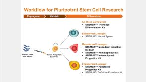
线上讲座STEMdiff™ Kits for Robust and Efficient Differentiation of hPSCs to Multiple Cell Types发布日期: 06/24/2016
20:24
线上讲座STEMdiff™ Kits for Robust and Efficient Differentiation of hPSCs to Multiple Cell Types发布日期: 06/24/2016 -
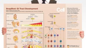 挂图SnapShot: GI Tract Development Overview of gastrointestinal tract specification signals and summary of pancreatic cellular hierarchy and cell markers
 -

-
-

 沪公网安备31010102008431号