
 EasySep™小鼠TIL(CD45)正选试剂盒


技术资料
-
-
-
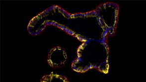
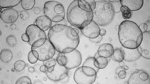
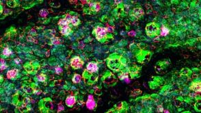
 实验方案Expansion of Hepatic Organoids via Single Cells Using HepatiCult™ Organoid Growth Medium
研究方向:
上皮细胞生物学,类器官
-
-
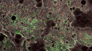
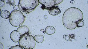
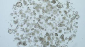
 实验方案Recovery and Expansion of Hepatic Organoids Using HepatiCult™ Organoid Growth Medium
研究方向:
上皮细胞生物学,类器官
-
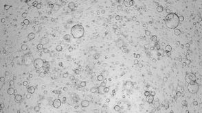
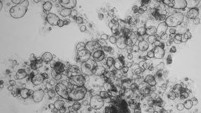
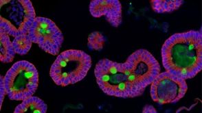
 实验方案Cryopreserving Hepatic Organoids Expanded in HepatiCult™ Organoid Growth Medium (Human)
研究方向:
上皮细胞生物学,类器官
-
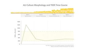
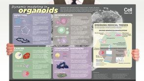 挂图Dynamic Modeling in Organoids Learn more about organoid applications for studying human health
 -
-
-

 沪公网安备31010102008431号