
 EasySep™小鼠TIL(CD45)正选试剂盒


技术资料
-
 科学海报Rapid, High-Efficiency Differentiation of Motor Neurons from Human Pluripotent Stem Cells
Conference:
FENS 2022
-
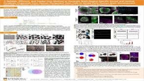
 科学海报Generation of a Glia-Midbrain Neuron Co-Culture System Derived From Human Pluripotent Stem Cells
Conference:
ISSCR 2022
-
 科学海报Efficient Differentiation of Human Pluripotent Stem Cells to Sensory Neurons
Conference:
ISSCR 2022
-
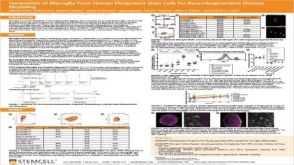 科学海报Generation of Microglia From Human Pluripotent Stem Cells for Neurodegenerative Disease Modeling
Conference:
ISSCR Toronto 2019
-
-


 沪公网安备31010102008431号