
 EasySep™小鼠TIL(CD45)正选试剂盒


技术资料
-
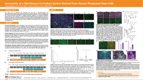 科学海报Generation of a Glia-Neuron Co-Culture System Derived From Human Pluripotent Stem Cells
Conference:
GLIA 2021
-
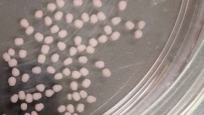 实验方案How to Dissociate 3D Neural Organoids into a Single-Cell Suspension
研究方向:
干细胞生物学,疾病建模,神经科学,类器官,药物发现和毒性检测,传染病
-
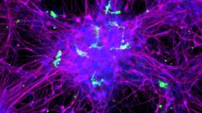 实验方案How to Co-Culture Human Pluripotent Stem Cell (hPSC)-Derived Forebrain Neurons and Microglia
研究方向:
免疫学,疾病建模,神经科学,药物发现和毒性检测,传染病
-

-
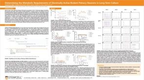 科学海报Determining the Metabolic Requirements of Electrically Active Rodent Primary Neurons in Long-Term Culture
Conference:
Society for Neuroscience Global Connectome 2021
发布日期: 03/17/2021 -
-
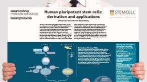 挂图Derivation and Applications of Human Pluripotent Stem Cells Overview of the derivation of human embryonic stem cells (hESCs) and induced pluripotent stem cells (iPSCs)发布日期: 11/26/2020


 沪公网安备31010102008431号