
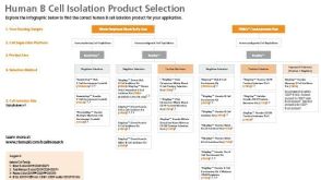
 EasySep™小鼠TIL(CD45)正选试剂盒


技术资料
-

-
-
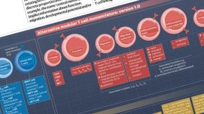 挂图T Cell Nomenclature: From Subsets to Modules A modular framework for classifying T cells by lineage, function, migration, differentiation, and antigen context.发布日期: 12/05/2025
 -
 实验方案How to Collect Plasma from Whole Blood Before Cell Isolation
研究方向:
免疫学,疾病建模,细胞外囊泡研究,细胞治疗开发,癌症研究
-
-

-
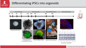
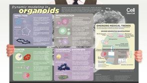 挂图Dynamic Modeling in Organoids Learn more about organoid applications for studying human health
 -


 沪公网安备31010102008431号