
 EasySep™小鼠TIL(CD45)正选试剂盒


技术资料
-
-
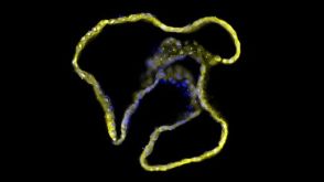
 研究综述The Predictive Power of Organoid-Based New Approach Methodologies in Drug Discovery
细胞类型:
上皮细胞,多能干细胞,肠道细胞,胰腺细胞,肾脏细胞,PSC衍生上皮细胞,PSC衍生肝细胞,呼吸道细胞
-
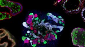
 技术公告Dendritic Cell/CD8+ T Cell Co-Culture to Assess Antigen-Specific T Cell Functionality
细胞类型:
CD8+ T细胞,T 细胞,单核细胞,树突状细胞(DCs)
-


 沪公网安备31010102008431号