Parmigiani A et al. (FEB 2011)
Human immunology 72 2 115--23
Interleukin-21 and cellular activation concurrently induce potent cytotoxic function and promote antiviral activity in human CD8 T cells.
Infection with human immunodeficiency virus (HIV)-1 induces a progressive deterioration of the immune system that ultimately leads to acquired immune deficiency syndrome (AIDS). Murine models indicate that the common γ-chain (γ(c))-sharing cytokine interleukin (IL)-21 and its receptor (IL-21R) play a crucial role in maintaining polyfunctional T cell responses during chronic viral infections. Therefore,we analyzed the ability of this cytokine to modulate the properties of human CD8 T cells in comparison with other γ(c)-sharing cytokines (IL-2,IL-7,and IL-15). CD8 T cells from healthy volunteers were stimulated in vitro via T cell receptor signals to mimic the heightened status of immune activation of HIV-infected patients. The administration of IL-21 upregulated cytotoxic effector function and the expression of the costimulatory molecule CD28. Notably,this outcome was not accompanied by increased cellular proliferation or activation. Moreover,IL-21 promoted antiviral activity while not inducing HIV-1 replication in vitro. Thus,IL-21 may be a favorable molecule for immunotherapy and a suitable vaccine adjuvant in HIV-infected individuals.
View Publication
产品号#:
15024
15064
15023
15063
15021
15061
产品名:
RosetteSep™ 人B细胞富集抗体混合物
RosetteSep™人B细胞富集抗体混合物
RosetteSep™ 人CD8+ T细胞富集抗体混合物
RosetteSep™人CD8+ T细胞富集抗体混合物
RosetteSep™人T细胞富集抗体混合物
RosetteSep™人T细胞富集抗体混合物
Sá et al. (JUN 2010)
Nature protocols 5 6 1033--41
Ex vivo T cell-based HIV suppression assay to evaluate HIV-specific CD8+ T-cell responses.
To advance T cell-based HIV vaccine development,it is necessary to evaluate the immune correlates of a protective CD8(+) T-cell response. We have developed an assay that assesses the capacity ex vivo of HIV-specific CD8(+) T cells to suppress HIV-1 infection of autologous CD4(+) T cells. This assay directly reflects the ultimate effector function of CD8(+) T cells,the elimination of infected cells,and accurately differentiates the effective CD8(+) T-cell response in spontaneous HIV controllers from ineffective responses in other patients. In this article,we describe all the steps from cell purification to assessment of viral replication by HIV-p24 ELISA and analysis,along with conditions for cell culturing,and how to choose the viral infectious dose that gives the most reliable results. We also depict the conditions of a rapid assay on the basis of flow cytometry analysis of intracellular HIV-Gag products. These procedures take 14-17 d when the p24 ELISA assay is used,or 6 d with the intracellular Gag assay.
View Publication
产品号#:
21000
20119
20155
19053
19053RF
20104
20124
产品名:
RoboSep™- S
RoboSep™ 吸头组件抛光剂
RoboSep™分选试管套装(9个塑料管+吸头保护器)
EasySep™人CD8+ T细胞富集试剂盒
RoboSep™ 人CD8+ T细胞富集试剂盒含滤芯吸头
RoboSep™ 缓冲液
RoboSep™ 缓冲液 (5X浓缩液)
Balkow S et al. (SEP 2010)
Blood 116 11 1885--94
LFA-1 activity state on dendritic cells regulates contact duration with T cells and promotes T-cell priming.
A key event in the successful induction of adaptive immune responses is the antigen-specific activation of T cells by dendritic cells (DCs). Although LFA-1 (lymphocyte function-associated antigen 1) on T cells is considered to be important for antigen-specific T-cell activation,the role for LFA-1 on DCs remains elusive. Using 2 different approaches to activate LFA-1 on DCs,either by deletion of the αL-integrin cytoplasmic GFFKR sequence or by silencing cytohesin-1-interacting protein,we now provide evidence that DCs are able to make use of active LFA-1 and can thereby control the contact duration with naive T cells. Enhanced duration of DC/T-cell interaction correlates inversely with antigen-specific T-cell proliferation,generation of T-helper 1 cells,and immune responses leading to delayed-type hypersensitivity. We could revert normal interaction time and T-cell proliferation to wild-type levels by inhibition of active LFA-1 on DCs. Our data further suggest that cytohesin-1-interacting protein might be responsible for controlling LFA-1 deactivation on mature DCs. In summary,our findings indicate that LFA-1 on DCs needs to be in an inactive state to ensure optimal T-cell activation and suggest that regulation of LFA-1 activity allows DCs to actively control antigen-driven T-cell proliferation and effective immune responses.
View Publication
产品号#:
21000
20119
20155
19752
19752RF
19753
19753RF
产品名:
RoboSep™- S
RoboSep™ 吸头组件抛光剂
RoboSep™分选试管套装(9个塑料管+吸头保护器)
Weiss L et al. (JUN 2010)
Proceedings of the National Academy of Sciences of the United States of America 107 23 10632--7
In vivo expansion of naive and activated CD4+CD25+FOXP3+ regulatory T cell populations in interleukin-2-treated HIV patients.
HIV-1 infection is characterized by a progressive decline in CD4(+) T cells leading to a state of profound immunodeficiency. IL-2 therapy has been shown to improve CD4(+) counts beyond that observed with antiretroviral therapy. Recent phase III trials revealed that despite a sustained increase in CD4(+) counts,IL-2-treated patients did not experience a better clinical outcome [Abrams D,et al. (2009) N Engl J Med 361(16):1548-1559]. To explain these disappointing results,we have studied phenotypic,functional,and molecular characteristics of CD4(+) T cell populations in IL-2-treated patients. We found that the principal effect of long-term IL-2 therapy was the expansion of two distinct CD4(+)CD25(+) T cell populations (CD4(+)CD25(lo)CD127(lo)FOXP3(+) and CD4(+)CD25(hi)CD127(lo)FOXP3(hi)) that shared phenotypic markers of Treg but could be distinguished by the levels of CD25 and FOXP3 expression. IL-2-expanded CD4(+)CD25(+) T cells suppressed proliferation of effector cells in vitro and had gene expression profiles similar to those of natural regulatory CD4(+)CD25(hi)FOXP3(+) T cells (Treg) from healthy donors,an immunosuppressive T cell subset critically important for the maintenance of self-tolerance. We propose that the sustained increase of the peripheral Treg pool in IL-2-treated HIV patients may account for the unexpected clinical observation that patients with the greatest expansion of CD4(+) T cells had a higher relative risk of clinical progression to AIDS.
View Publication
产品号#:
15022
15062
产品名:
RosetteSep™人CD4+ T细胞富集抗体混合物
RosetteSep™人CD4+ T细胞富集抗体混合物
Vieillard V et al. (AUG 2005)
Proceedings of the National Academy of Sciences 102 31 10981--86
NK cytotoxicity against CD4+ T cells during HIV-1 infection: A gp41 peptide induces the expression of an NKp44 ligand
HIV infection leads to a state of chronic immune activation and progressive deterioration in immune function,manifested most recognizably by the progressive depletion of CD4+ T cells. A substantial percentage of natural killer (NK) cells from patients with HIV infection are activated and express the natural cytotoxicity receptor (NCR) NKp44. Here we show that a cellular ligand for NKp44 (NKp44L) is expressed during HIV-1 infection and is correlated with both the progression of CD4+ T cell depletion and the increase of viral load. CD4+ T cells expressing this ligand are highly sensitive to the NK lysis activity mediated by NKp44+ NK cells. The expression of NKp44L is induced by the linear motif NH2-SWSNKS-COOH of the HIV-1 envelope gp41 protein. This highly conserved motif appears critical to the sharp increase in NK lysis of CD4+ T cells from HIV-infected patients. These studies strongly suggest that induction of NKp44L plays a key role in the lysis of CD4+ T cells by activated NK cells in HIV infection and consequently provide a framework for considering how HIV-1 may use NK cell immune surveillance to trigger CD4+ T cells. Understanding this mechanism may help to develop future therapeutic strategies and vaccines against HIV-1 infection.
View Publication
产品号#:
03800
03801
03802
03803
03804
03805
03806
05150
15021
15061
产品名:
ClonaCell™-HY 杂交瘤试剂盒
ClonaCell™-HY Medium
ClonaCell™-HY Medium
ClonaCell™-HY Medium
ClonaCell™-HY Medium
ClonaCell™-HY Medium
ClonaCell™-HY PEG (融合)
MyeloCult™H5100
RosetteSep™人T细胞富集抗体混合物
RosetteSep™人T细胞富集抗体混合物
Abdelwahab SF et al. (DEC 2003)
Proceedings of the National Academy of Sciences of the United States of America 100 25 15006--10
HIV-1-suppressive factors are secreted by CD4+ T cells during primary immune responses.
CD4+ T cells are required for immunity against many viral infections,including HIV-1 where a positive correlation has been observed between strong recall responses and low HIV-1 viral loads. Some HIV-1-specific CD4+ T cells are preferentially infected with HIV-1,whereas others escape infection by unknown mechanisms. One possibility is that some CD4+ T cells are protected from infection by the secretion of soluble HIV-suppressive factors,although it is not known whether these factors are produced during primary antigen-specific responses. Here,we show that soluble suppressive factors are produced against CXCR4 and CCR5 isolates of HIV-1 during the primary immune response of human CD4+ T cells. This activity requires antigenic stimulation of naïve CD4+ T cells. One anti-CXCR4 factor is macrophage-derived chemokine (chemokine ligand 22,CCL22),and anti-CCR5 factors include macrophage inflammatory protein-1 alpha (CCL3),macrophage inflammatory protein-1 beta (CCL4),and RANTES (regulated upon activation of normal T cells expressed and secreted) (CCL5). Intracellular staining confirms that CD3+CD4+ T cells are the source of the prototype HIV-1-inhibiting chemokines CCL22 and CCL4. These results show that CD4+ T cells secrete an evolving HIV-1-suppressive activity during the primary immune response and that this activity is comprised primarily of CC chemokines. The data also suggest that production of such factors should be considered in the design of vaccines against HIV-1 and as a mechanism whereby the host can control infections with this virus.
View Publication
产品号#:
09500
09600
09650
19155
19155RF
产品名:
BIT 9500血清替代物
StemSpan™ SFEM
StemSpan™ SFEM
Jones DC et al. (JUL 2003)
Journal of immunology 171 1 196--203
Peroxisome proliferator-activated receptor alpha negatively regulates T-bet transcription through suppression of p38 mitogen-activated protein kinase activation.
Expression of the nuclear hormone receptor peroxisome proliferator-activated receptor alpha (PPARalpha) in resting lymphocytes was recently established,although the physiologic role(s) played by this nuclear hormone receptor in these cell types remains unresolved. In this study,we used CD4(+) T cells isolated from PPARalpha(-/-) and wild-type mice,as well as cell lines that constitutively express PPARalpha,in experiments designed to evaluate the role of this hormone receptor in the regulation of T cell function. We report that activated CD4(+) T cells lacking PPARalpha produce increased levels of IFN-gamma,but significantly lower levels of IL-2 when compared with activated wild-type CD4(+) T cells. Furthermore,we demonstrate that PPARalpha regulates the expression of these cytokines by CD4(+) T cells in part,through its ability to negatively regulate the transcription of T-bet. The induction of T-bet expression in CD4(+) T cells was determined to be positively influenced by p38 mitogen-activated protein (MAP) kinase activation,and the presence of unliganded PPARalpha effectively suppressed the phosphorylation of p38 MAP kinase. The activation of PPARalpha with highly specific ligands relaxed its capacity to suppress p38 MAP kinase phosphorylation and promoted T-bet expression. These results demonstrate a novel DNA-binding independent and agonist-controlled regulatory influence by the nuclear hormone receptor PPARalpha.
View Publication
产品号#:
03814
产品名:
ClonaCell™-TCS培养基
Schlecht G et al. (OCT 2001)
Journal of immunology (Baltimore,Md. : 1950) 167 8 4215--21
Induction of CTL and nonpolarized Th cell responses by CD8alpha(+) and CD8alpha(-) dendritic cells.
Two distinct dendritic cell (DC) subpopulations have been evidenced in mice on the basis of their differential CD8alpha expression and their localization in lymphoid organs. Several reports suggest that CD8alpha(+) and CD8alpha(-) DC subsets could be functionally different. In this study,using a panel of MHC class I- and/or class II-restricted peptides,we analyzed CD4(+) and CD8(+) T cell responses obtained after i.v. injection of freshly purified peptide-pulsed DC subsets. First,we showed that both DC subsets efficiently induce specific CTL responses and Th1 cytokine production in the absence of CD4(+) T cell priming. Second,we showed that in vivo activation of CD4(+) T cells by CD8alpha(+) or CD8alpha(-) DC,injected i.v.,leads to a nonpolarized Th response with production of both Th1 and Th2 cytokines. The CD8alpha(-) subset induced a higher production of Th2 cytokines such as IL-4 and IL-10 than the CD8alpha(+) subset. However,IL-5 was produced by CD4(+) T cells activated by both DC subsets. When both CD4(+) and CD8(+) T cells were primed by DC injected i.v.,a similar pattern of cytokines was observed,but,under these conditions,Th1 cytokines were mainly produced by CD8(+) T cells,while Th2 cytokines were produced by CD4(+) T cells. Thus,this study clearly shows that CD4(+) T cell responses do not influence the development of specific CD8(+) T cell cytotoxic responses induced either by CD8alpha(+) or CD8alpha(-) DC subsets.
View Publication

 EasySep™小鼠TIL(CD45)正选试剂盒


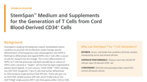 技术公告StemSpan™ Medium and Supplements for the Generation of T Cells from Cord Blood-Derived CD34+ Cells


 沪公网安备31010102008431号