若您需要咨询产品或有任何技术问题,请通过官方电话 400 885 9050 或邮箱 info.cn@stemcell.com 与我们联系

EasySep™ HLA嵌合全血CD66b正选试剂盒

从全血中免疫磁珠正选人CD66+细胞

产品号 #(选择产品)

产品号 #17882_C

从全血中免疫磁珠正选人CD66+细胞

产品优势

  • 操作简单、快速
  • 纯度高达99%
  • 无需分离柱

产品组分包括

  • EasySep™ HLA嵌合全血CD66b正选试剂盒(产品号 #17882)
    • EasySep™ HLA嵌合全血CD66b正选抗体混合物,3 x 1 mL
    • EasySep™ Dextran RapidSpheres™ 50102磁珠,3 x 1 mL
    • EasySep™ 10X浓缩红细胞裂解试剂,10 mL(产品号# 20110)    
  • RoboSep™ HLA嵌合全血CD66b正选试剂盒(含过滤枪头)(产品 号 #17882RF)
    • EasySep™ HLA嵌合全血CD66b正选抗体混合物,3 x 1 mL    
    • EasySep™ Dextran RapidSpheres™ 50102磁珠,3 x 1 mL
    • EasySep™ 10X浓缩红细胞裂解试剂,10 mL(产品号# 20110)     RoboSep™ 缓冲液(产品号 #20104)x 2
    • RoboSep™ 过滤枪头(产品号 #20125)x 2
New look, same high quality and support! You may notice that your instrument or reagent packaging looks slightly different from images displayed on the website, or from previous orders. We are updating our look but rest assured, the products themselves and how you should use them have not changed. Learn more
专为您的实验方案打造的产品

总览

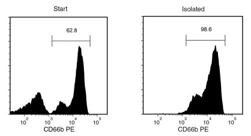
使用EasySep™ HLA嵌合全血CD66b正选试剂盒,从新鲜人全血样本中通过免疫磁珠正选分选高纯度人CD66b+细胞。EasySep™技术结合单克隆抗体的特异性和无柱磁珠系统的简便性,已在发表的研究中广泛应用超过20年。

在此EasySep™正选流程中,目的细胞通过被CD66b的抗体复合物与磁珠标记,并通过 EasySep™ 磁极进行无柱分选 ,只需倾倒非目标细胞即可,而目标细胞保留在管中。分选后的CD66b+ 细胞可立即用于下游应用,例如流式 、培养、或DNA/RNA提取,用于谱系特异性嵌合分析。CD66b抗原在成熟粒细胞上表达。

本 产品可替代EasySep™人全血 CD66b正选试剂盒 (产品号 #18682) 以进行更快地细胞分选。

深入了解免疫磁珠EasySep™技术的工作原理,或如何通过RoboSep™实现全自动化的免疫磁珠细胞分选,以节省时间并提升实验室通量。探索更多为您的实验流程优化的产品,包括细胞表征、冷存等相关产品。

磁体兼容性
• “The Big Easy” EasySep™ Magnet (Catalog #18001) • EasyEights™ EasySep™ Magnet (Catalog #18103) • RoboSep™-S (Catalog #21000)
 
亚型
细胞分选试剂盒
 
细胞类型
粒细胞及其亚群
 
种属

 
样本来源
Whole Blood
 
筛选方法
Positive
 
应用
细胞分选
 
品牌
EasySep,RoboSep
 
研究领域
嵌合体,免疫
 

实验数据

Typical EasySep™ HLA Chimerism Whole Blood CD66b Positive Selection Profile

Figure 1. Typical EasySep™ HLA Chimerism Whole Blood CD66b Positive Selection Profile

Starting with whole blood, the CD66b+ cell content of the isolated fraction is typically 98.0 ± 0.8% (mean ± SD) using “The Big Easy” EasySep™ Magnet. In the above example, the purities of the start and final isolated fractions are 62.8% and 98.6%, respectively.

产品说明书及文档

请在《产品说明书》中查找相关支持信息和使用说明,或浏览下方更多实验方案。

Document Type
Product Name
Catalog #
Lot #
Language
Catalog #
17882RF
Lot #
1000131008 or higher
Language
English
Catalog #
17882
Lot #
1000131008 or higher
Language
English
Document Type
Safety Data Sheet 1
Catalog #
17882RF
Lot #
All
Language
English
Document Type
Safety Data Sheet 2
Catalog #
17882RF
Lot #
All
Language
English
Document Type
Safety Data Sheet 3
Catalog #
17882RF
Lot #
All
Language
English
Document Type
Safety Data Sheet 4
Catalog #
17882RF
Lot #
All
Language
English
Document Type
Safety Data Sheet 1
Catalog #
17882
Lot #
All
Language
English
Document Type
Safety Data Sheet 2
Catalog #
17882
Lot #
All
Language
English
Document Type
Safety Data Sheet 3
Catalog #
17882
Lot #
All
Language
English

应用领域

本产品专为以下研究领域设计,适用于工作流程中的高亮阶段。探索这些工作流程,了解更多我们为各研究领域提供的其他配套产品。

相关材料与文献

技术资料 (11)

文献 (3)

Single-cell analysis of menstrual endometrial tissues defines phenotypes associated with endometriosis. A. J. Shih et al. BMC medicine 2022 sep

Abstract

BACKGROUND Endometriosis is a common, complex disorder which is underrecognized and subject to prolonged delays in diagnosis. It is accompanied by significant changes in the eutopic endometrial lining. METHODS We have undertaken the first single-cell RNA-sequencing (scRNA-Seq) comparison of endometrial tissues in freshly collected menstrual effluent (ME) from 33 subjects, including confirmed endometriosis patients (cases) and controls as well as symptomatic subjects (who have chronic symptoms suggestive of endometriosis but have not been diagnosed). RESULTS We identify a unique subcluster of proliferating uterine natural killer (uNK) cells in ME-tissues from controls that is almost absent from endometriosis cases, along with a striking reduction of total uNK cells in the ME of cases (p??<??10-16). In addition, an IGFBP1+ decidualized subset of endometrial stromal cells are abundant in the shed endometrium of controls when compared to cases (p??<??10-16) confirming findings of compromised decidualization of cultured stromal cells from cases. By contrast, endometrial stromal cells from cases are enriched in cells expressing pro-inflammatory and senescent phenotypes. An enrichment of B cells in the cases (p??=??5.8???—??10-6) raises the possibility that some may have chronic endometritis, a disorder which predisposes to endometriosis. CONCLUSIONS We propose that characterization of endometrial tissues in ME will provide an effective screening tool for identifying endometriosis in patients with chronic symptoms suggestive of this disorder. This constitutes a major advance, since delayed diagnosis for many years is a major clinical problem in the evaluation of these patients. Comprehensive analysis of ME is expected to lead to new diagnostic and therapeutic approaches to endometriosis and other associated reproductive disorders such as female infertility.
IL-1 and IL-1ra are key regulators of the inflammatory response to RNA vaccines. S. Tahtinen et al. Nature immunology 2022 apr

Abstract

The use of lipid-formulated RNA vaccines for cancer or COVID-19 is associated with dose-limiting systemic inflammatory responses in humans that were not predicted from preclinical studies. Here, we show that the 'interleukin 1 (IL-1)-interleukin 1 receptor antagonist (IL-1ra)' axis regulates vaccine-mediated systemic inflammation in a host-specific manner. In human immune cells, RNA vaccines induce production of IL-1 cytokines, predominantly IL-1$\beta$, which is dependent on both the RNA and lipid formulation. IL-1 in turn triggers the induction of the broad spectrum of pro-inflammatory cytokines (including IL-6). Unlike humans, murine leukocytes respond to RNA vaccines by upregulating anti-inflammatory IL-1ra relative to IL-1 (predominantly IL-1$\alpha$), protecting mice from cytokine-mediated toxicities at >1,000-fold higher vaccine doses. Thus, the IL-1 pathway plays a key role in triggering RNA vaccine-associated innate signaling, an effect that was unexpectedly amplified by certain lipids used in vaccine formulations incorporating N1-methyl-pseudouridine-modified RNA to reduce activation of Toll-like receptor signaling.
Tumor-Infiltrating PD-L1+ Neutrophils Induced by GM-CSF Suppress T Cell Function in Laryngeal Squamous Cell Carcinoma and Predict Unfavorable Prognosis. D. Tang et al. Journal of inflammation research 2022

Abstract

PURPOSE Chronic inflammation contributes to tumor initiation, progression, and immune escape. Neutrophils are the major component of inflammatory response and participate in the tumorigenesis process. However, compared to other immune cells in the tumor microenvironment of laryngeal squamous cell carcinoma (LSCC), neutrophils, especially the tumor-associated neutrophils (TANs), have not yet been comprehensively explored. The mechanism for regulating the crosstalk between TANs and tumor cells still remains unclear. MATERIALS AND METHODS The distribution profiles and phenotypic features of neutrophils and other inflammatory immune cell populations from a large LSCC patient cohort were systemically analyzed. Co-culturing of peripheral blood associated neutrophils (PANs) and TANs with PBMCs was performed, and the immunosuppression effect on T-cells was examined. RESULTS LSCC microenvironment is highly inflammatory with remarkable TANs infiltration, which is often associated with unfavorable prognosis and advanced clinical stage. We find that TANs in LSCC display morphologically immature and lower apoptosis, exhibit distinctively immunosuppressive phenotype of high PD-L1, and suppress CD8+ T lymphocytes proliferation and activation. We subsequently discover that PD-L1+TANs induced by LSCC-derived GM-CSF potently impair CD8+ T-cells proliferation and cytokines production function, which are partially blocked by a PD-L1-neutralizing antibody. Clinical data further support GM-CSF as an unfavorable prognostic biomarker and reveal a potential association with inflammatory immune cell infiltration, in particular neutrophils. CONCLUSION Tumor-infiltrating PD-L1+ neutrophils induced by LSCC-derived GM-CSF suppress T cell proliferation and activation in the inflammatory microenvironment of LSCC and predict unfavorable prognosis. These TANs cripple antitumor T cell immunity and promote tumor progression. Our findings provide a basis for targeting PD-L1+TANs or GM-CSF as a new immunotherapeutic strategy for LSCC.

更多信息

更多信息
种属 Human
Magnet Compatibility • “The Big Easy” EasySep™ Magnet (Catalog #18001) • EasyEights™ EasySep™ Magnet (Catalog #18103) • RoboSep™-S (Catalog #21000)
样本来源 Whole Blood
Selection Method Positive
标记抗体
质量保证:

产品仅供研究使用,不用于针对人或动物的诊断或治疗。 欲获悉更多关于STEMCELL的质控信息,请访问 STEMCELL.CN/COMPLIANCE.
Copyright © 2025 by STEMCELL Technologies. All rights reserved.