
 EasySep™小鼠TIL(CD45)正选试剂盒


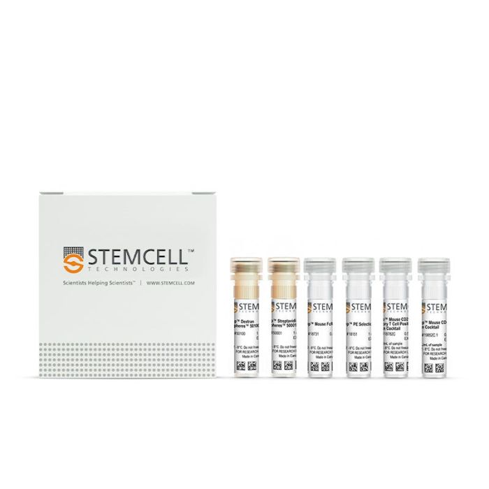
产品号 #18783_C
通过免疫磁珠正选分离小鼠CD4+CD25+调节性T细胞
若您需要咨询产品或有任何技术问题,请通过官方电话 400 885 9050 或邮箱 info.cn@stemcell.com 与我们联系。
通过免疫磁珠正选分离小鼠CD4+CD25+调节性T细胞
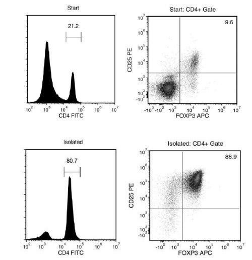
使用EasySep™小鼠CD4+CD25+调节性T细胞分选试剂盒II,通过简单的两步法(首先进行免疫磁珠负选,然后使用正选),即可轻松从小鼠脾细胞或其他单细胞悬液样本中分离出高纯度的小鼠CD4+CD25+调节性T细胞(Tregs)。EasySep™在已发表的研究中被广泛使用了20多年,它结合了单克隆抗体的特异性和无柱磁珠系统的简便性。
在此EasySep™细胞分选过程中,首先使用EasySep™小鼠CD4+ T细胞分选抗体混合物通过负选富集CD4+ T细胞。然后使用EasySep™小鼠CD25调节性T细胞正选抗体混合物从预富集的细胞中正选分离出CD25+细胞。完成磁珠分选后,获得的CD4+CD25+ Tregs即可用于下游应用,例如流式细胞术、培养、DNA/RNA提取。
了解有关EasySep™免疫磁珠技术的工作原理或如何使用RoboSep™进行全自动免疫磁珠细胞分选的更多信息。探索针对您的工作流程进行优化的其他产品,包括培养基、添加物、抗体等。
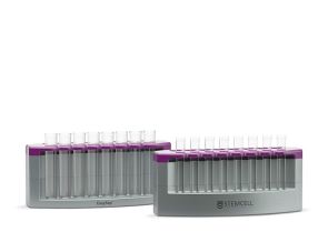
磁极兼容性
• EasySep™磁极(产品号 #18000)
• “The Big Easy” EasySep™磁极(产品号 #18001)
• EasyEights™ EasySep™磁极(产品号 #18103)
• RoboSep™-S(产品号 #21000)
分类
细胞分选试剂盒
细胞类型
T 细胞,T 细胞,调节性细胞
种属
小鼠
样本来源
脾脏
品牌
EasySep,RoboSep
研究领域
免疫
请在《产品说明书》中查找相关支持信息和使用说明,或浏览下方更多实验方案。
本产品专为以下研究领域设计,适用于工作流程中的高亮阶段。探索这些工作流程,了解更多我们为各研究领域提供的其他配套产品。
| 物种 | 小鼠 |
|---|---|
| Magnet Compatibility | • EasySep™ Magnet (Catalog #18000) • “The Big Easy” EasySep™ Magnet (Catalog #18001) • EasyEights™ Magnet (Catalog #18103) • RoboSep™-S (Catalog #21000) |
| 样本来源 | 脾脏 |
<p>对来源于小鼠脾细胞或其他单细胞悬液的 CD25⁺ 调节性 T 细胞(Tregs)进行免疫磁珠正选分离</p>
<p>通过免疫磁珠负选获得无磁珠标记的小鼠T细胞</p>
<p>抗小鼠CD4的大鼠Monoclonal lgG2a抗体</p>

抗小鼠CD4的大鼠Monoclonal IgG2b抗体
在线联系


 沪公网安备31010102008431号