
 EasySep™小鼠TIL(CD45)正选试剂盒


技术资料
-
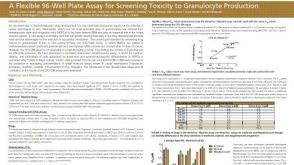 科学海报A Flexible 96-Well Plate Assay for Screening Toxicity to Granulocyte Production
Conference:
SOT 2015
-
-
-
 技术窍门CFU检测中造血祖细胞的培养与分析
 -
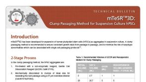
 技术公告StemSpan™ Medium and Supplements for the Generation of T Cells from Cord Blood-Derived CD34+ Cells
细胞类型:
CD4+ T细胞,CD8+ T细胞,T 细胞,造血干/祖细胞
-
 技术窍门催化成熟扩增培养的红系祖细胞
 -
-
-
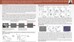 科学海报Generation and Characterization of a Homogenous Population of Early Mesoderm Cells Using STEMdiff Mesoderm Induction Medium
Conference:
ISSCR 2015; TMM 2015


 沪公网安备31010102008431号