
 EasySep™小鼠TIL(CD45)正选试剂盒


技术资料
-
-
-
-
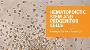 产品手册Your Ideas. Our Tools. 为您提供造血干细胞和祖细胞研究 各个步骤的相关产品
品牌:
ALDEFLUOR,MethoCult,MyeloCult,STEMdiff,STEMvision,SmartDish,StemSpan,ThawSTAR
-
-
-


 沪公网安备31010102008431号