
 EasySep™小鼠TIL(CD45)正选试剂盒


产品号 #100-0900_C
用于将人胚胎干细胞(ES细胞)或诱导多能干细胞(iPS细胞)分化为巨核细胞和血小板。
若您需要咨询产品或有任何技术问题,请通过官方电话 400 885 9050 或邮箱 info.cn@stemcell.com 与我们联系。
用于将人胚胎干细胞(ES细胞)或诱导多能干细胞(iPS细胞)分化为巨核细胞和血小板。
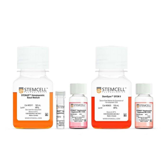
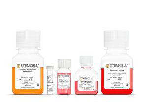
STEMdiff™ 巨核细胞试剂盒专为在无血清、无饲养层的条件下,将人类胚胎干细胞(ES细胞)和诱导多能干细胞(iPS细胞)分化为具有多倍体、可释放血小板并表达 CD41a 和 CD42b 的巨核细胞而设计。本试剂盒采用一个简单的二维、为期17天的分化方案,包含两个阶段。
在前 12 天,将 STEMdiff™ 造血补充剂 A 和补充剂 MK1 添加到基础培养基中,以诱导细胞向巨核细胞倾向的造血祖细胞分化。在此阶段结束时,可从培养上清液中轻松收获造血祖细胞,并使用补充剂 MK2 和 StemSpan™ SFEM II 继续培养 5 天,使其进一步分化为成熟的巨核细胞。
在方案结束时(第17天),细胞通常可扩增至初始数量的约405倍±54倍(95% 置信区间),平均约有71%±3.3%(95% 置信区间)的细胞共表达CD41a和CD42b。
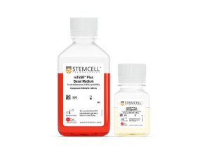
STEMdiff™ 巨核细胞试剂盒已针对在以下培养体系中维持的 ES/iPS 细胞进行了优化:mTeSR™1(产品号 85850)、mTeSR™ Plus(产品号 100-0276)或 TeSR™-AOF(产品号 100-0401)。
分类
专用培养基
细胞类型
造血细胞,PSC衍生,造血干/祖细胞,巨核细胞
应用
分化
品牌
STEMdiff
研究领域
疾病建模,药物发现和毒理检测,干细胞生物学,细胞治疗开发
制剂类别
无血清
请在《产品说明书》中查找相关支持信息和使用说明,或浏览下方更多实验方案。
本产品专为以下研究领域设计,适用于工作流程中的高亮阶段。探索这些工作流程,了解更多我们为各研究领域提供的其他配套产品。
| 配方 | 无血清 |
|---|
含重组细胞因子的无血清甲基纤维素培养基,适用于人 ES 和 iPS 细胞衍生细胞
用于将人ES细胞或iPS细胞分化为造血祖细胞
用于将人 ES 细胞或 iPS 细胞分化为红系祖细胞

<p>cGMP标准、无饲养层的hESC和iPSC维持培养基</p>
在线联系


 沪公网安备31010102008431号