
 EasySep™小鼠TIL(CD45)正选试剂盒


产品号 #100-1245_C
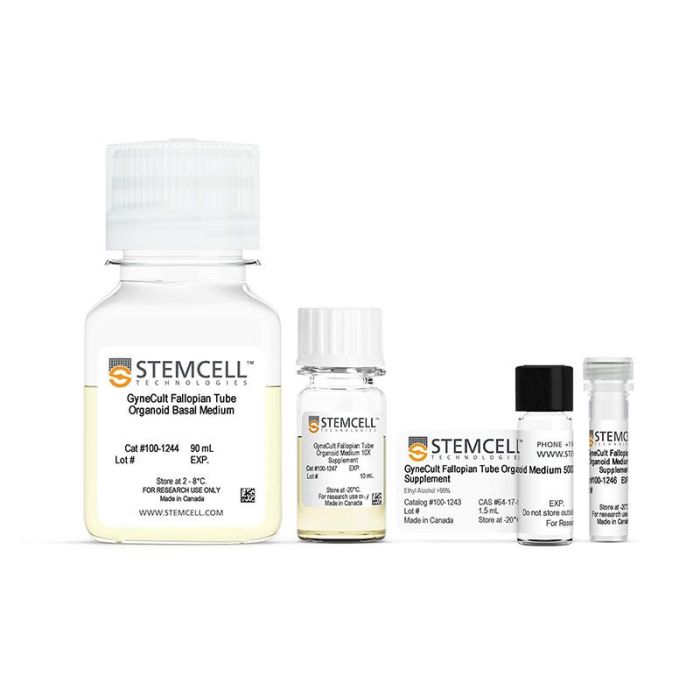
用于扩增和分化人输卵管类器官的无血清、支持性激素调节的试剂盒
若您需要咨询产品或有任何技术问题,请通过官方电话 400 885 9050 或邮箱 info.cn@stemcell.com 与我们联系。
用于扩增和分化人输卵管类器官的无血清、支持性激素调节的试剂盒
用于扩增和分化人输卵管类器官的无血清、支持性激素调节的试剂盒
生成成熟的输卵管(fallopian tube, FT)类器官,呈现出与体内相似的纤毛细胞和分泌细胞的代表性组合。GyneCult™ 输卵管类器官培养基(FTOM)在功能性输卵管类器官培养中表现优于自配体系(DIY 配方),能够更好地支持相关细胞类型在生理比例下的生长。
由GyneCult™FTOM生成的类器官为女性生殖生物学研究提供了一个强大而通用的系统。通过该优化的工作流程,可以直接接种成3D培养从而节省时间和劳动力,并在第10天即可获得可传代的输卵管类器官。GyneCult™ FTOM 是一款无血清、性激素调节模块化的试剂盒;通过不添加 5000X 补充剂,用户可根据需要灵活调节激素水平,同时为特定应用提供一致、可重复的实验结果。
输卵管类器官培养的应用包括研究生育机制、激素动力学、月经及排卵后周期,以及卵巢癌。借助 GyneCult™ FTOM,可培养部分高等级浆液性卵巢癌(HGSOC)类器官,用于卵巢癌研究,例如评估输卵管上皮细胞的肿瘤发生能力,或用于卵巢癌化疗药物的筛选。
分类
专用培养基
细胞类型
上皮细胞
应用
细胞培养,分化,扩增,类器官培养
品牌
GyneCult
研究领域
癌症,疾病建模,药物发现和毒理检测,上皮细胞研究,类器官
制剂类别
无血清
请在《产品说明书》中查找相关支持信息和使用说明,或浏览下方更多实验方案。
本产品专为以下研究领域设计,适用于工作流程中的高亮阶段。探索这些工作流程,了解更多我们为各研究领域提供的其他配套产品。
| 配方 | 无血清 |
|---|
用于在气液界面培养的人呼吸道上皮细胞的无血清和无BPE培养基
用于 CFU 检测人乳腺上皮细胞的培养

用于培养人、小鼠和大鼠上皮干细胞
在线联系


 沪公网安备31010102008431号