
 EasySep™小鼠TIL(CD45)正选试剂盒


技术资料
-
-

-
-
-
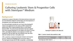 技术公告Culturing Leukemic Stem & Progenitor Cells with StemSpan™ Medium
细胞类型:
白血病/淋巴瘤细胞
发布日期: 11/14/2019 -
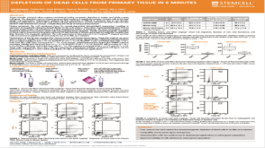
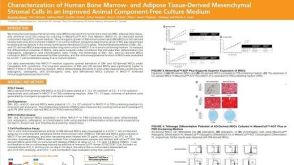 科学海报Characterization of Human Bone Marrow- and Adipose Tissue-Derived Mesenchymal Stromal Cells in an Improved Animal Component-Free Culture Medium
Conference:
ISSCR 2019
发布日期: 11/12/2019


 沪公网安备31010102008431号